Copyright
©2006 Baishideng Publishing Group Co.
World J Gastroenterol. Aug 21, 2006; 12(31): 5091-5093
Published online Aug 21, 2006. doi: 10.3748/wjg.v12.i31.5091
Published online Aug 21, 2006. doi: 10.3748/wjg.v12.i31.5091
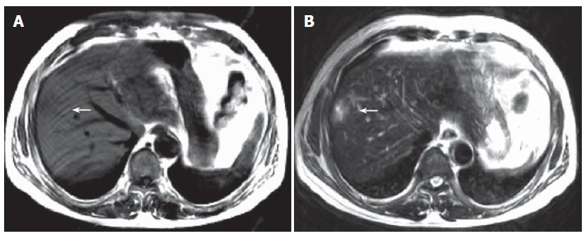
Figure 1 A: Between the V and VIIIsegments, the right hepatic lobe near the surface has an abnormal signal which seems ellipse.
In T1WI, its center shows long T1 and slight long T1 intensity in row; B: In T2WI, its center shows long T2 and slightly long T2 intensity in a row. It shows slight mass effect.
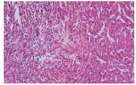
Figure 2 Pathologic section shows hepatic cell necrosis.
Vasodilatation and infiltration of inflammatory cells are seen at the junction of necrosis and normal tissues (HE × 100).
- Citation: Deng YG, Zhao ZS, Wang M, Su SO, Yao XX. Diabetes mellitus with hepatic infarction: A case report with literature review. World J Gastroenterol 2006; 12(31): 5091-5093
- URL: https://www.wjgnet.com/1007-9327/full/v12/i31/5091.htm
- DOI: https://dx.doi.org/10.3748/wjg.v12.i31.5091


