Copyright
©2006 Baishideng Publishing Group Co.
World J Gastroenterol. Aug 21, 2006; 12(31): 5084-5086
Published online Aug 21, 2006. doi: 10.3748/wjg.v12.i31.5084
Published online Aug 21, 2006. doi: 10.3748/wjg.v12.i31.5084
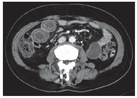
Figure 1 Abdominal computed tomography on admission revealed layering appearance of bowel wall and luminal dilatation from the distal jejunum to the pelvic ileal loop.
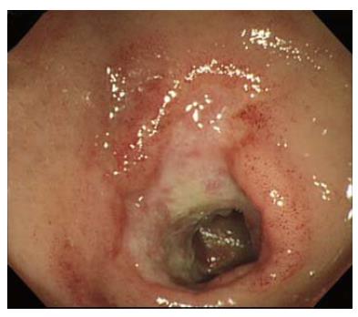
Figure 2 Colonoscopy, performed on admission, revealed scattered regions of geographic ulcers and hyperemic edematous mucosa on the terminal ileum.
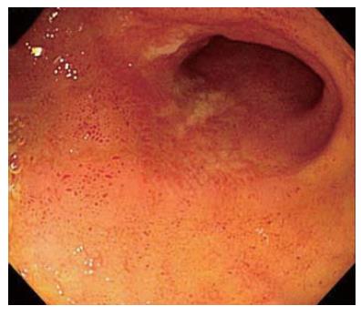
Figure 3 Follow up colonoscopy was performed.
The previously noted ulcers and hyperemic edematous lesion were improved.
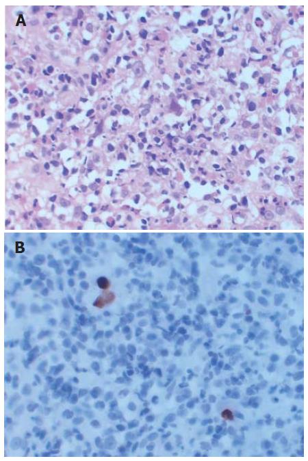
Figure 4 Pathologic findings.
A: Enlargement of infected cells and intranuclear inclusions were noted (HE stain, × 100); B: Immunohistochemical stain for CMV showed scattered positive cells.
- Citation: Ryu KH, Yi SY. Cytomegalovirus ileitis in an immunocompetent elderly adult. World J Gastroenterol 2006; 12(31): 5084-5086
- URL: https://www.wjgnet.com/1007-9327/full/v12/i31/5084.htm
- DOI: https://dx.doi.org/10.3748/wjg.v12.i31.5084


