Copyright
©2006 Baishideng Publishing Group Co.
World J Gastroenterol. Aug 21, 2006; 12(31): 5075-5077
Published online Aug 21, 2006. doi: 10.3748/wjg.v12.i31.5075
Published online Aug 21, 2006. doi: 10.3748/wjg.v12.i31.5075
Figure 1 Computed tomography (CT) scan showing a regular contoured 4 cm x 3 cm lesion with fatty density localised in midportion of ascending colon causing a luminal narrowing defect (A), double contrast barium enema showing a filling defect of the protruded polipoid lesion at the hepatic flexura of colon (B), and appearance of the colonic submucosal lipoma during the operation (C).
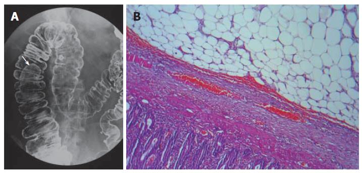
Figure 2 A mass lesion located in the hepatic flexura causing filling defect shown by double contrast enema (A), resected biopsy specimen showing a submucosal benign lipoma composed of mature lipocytes by hematoxylin and eosin staining (B) (x 400).
Figure 3 Colonoscopic appearance of a broad- based giant mass acquiring spontaneous hemorrhagic areas and obstructing more than 75% of the lumen of sigmoid colon (A); abdominal computed tomography scans showing the diffuse thickening of sigmoid colon wall, invagination and a distal intraluminal giant mass with fat density with a size of approximately 6 cm x 7 cm (B); macroscopic appearance of the giant sigmoid colon lipoma (C).
- Citation: Tascilar O, Cakmak GK, Gün BD, Uçan BH, Balbaloglu H, Cesur A, Emre AU, Comert M, Erdem LO, Aydemir S. Clinical evaluation of submucosal colonic lipomas: Decision making. World J Gastroenterol 2006; 12(31): 5075-5077
- URL: https://www.wjgnet.com/1007-9327/full/v12/i31/5075.htm
- DOI: https://dx.doi.org/10.3748/wjg.v12.i31.5075


