Copyright
©2006 Baishideng Publishing Group Co.
World J Gastroenterol. Jan 21, 2006; 12(3): 363-371
Published online Jan 21, 2006. doi: 10.3748/wjg.v12.i3.363
Published online Jan 21, 2006. doi: 10.3748/wjg.v12.i3.363
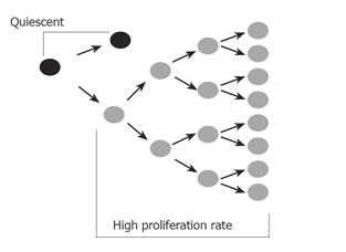
Figure 1 A proposed model for the cancer stem cell.
The cancer stem cell replicates itself asymmetrically, thus maintaining one daughter stem cell identical to itself. This remains in a relatively quiescent state. The asymmetric division also produces another daughter cell with a high proliferative rate which rapidly divides to sustain the tumor mass.
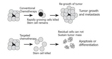
Figure 2 Conventional versus stem cell-targeted chemotherapy.
Conventional chemotherapy and radiotherapy targets rapidly dividing cells, and may shrink tumor mass substantially. However, the stem cell (gray), which is relatively quiescent, is not affected. Regrowth of tumor from surviving stem cell leads to regrowth of tumor and treatment failure. Chemotherapy targeted at the stem cell would remove the source of new cell growth, and allow residual cells within the tumor to be targeted with chemotherapy, differentiating agents or therapy aimed at inducing apoptosis, thus successfully eliminating the tumor.
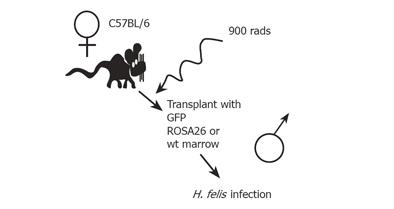
Figure 3 An experimental mouse model for bone marrow transplantation and H.
felis-induced gastric carcinoma.
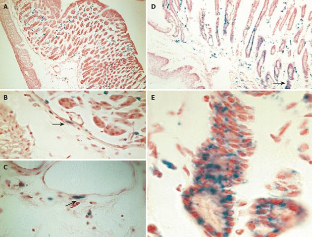
Figure 4 Engraftment of donor-derived ROSA-26 marrow by x-gal staining.
A: Mice transplanted with ROSA 26 marrow and infected with H. felis for 4 wk had donor-derived leukocytes (blue) infiltrating the gastric mucosa, and no engraftment into gland structures. B and C: A higher power view reveals myocytes and myofibroblasts in the submucosal tissue adjacent to vascular structures (arrows). D: After 30 wk of infection, marked architectural distortion is seen with antralization and appearance of metaplastic glands. Entire gland structures are derived from donor marrow (blue staining). Gland shown in panel D (arrow) is shown at higher power in E.
Figure 5 Immunohistochemistry for bacterial beta-galactosidase confirms uniform signal in gastrointestinal neoplasia.
Mice developed severe dysplasia and intraepithelial neoplasia derived from donor marrow, 12-15 mo after infection with H. felis (A) and (B). Immunohistochemistry for bacterial beta-galactosidase demonstrates cytoplasmic staining in dysplastic glands. A population of adipocytes in the submucosa are also stained for beta-galactosidase (arrow).
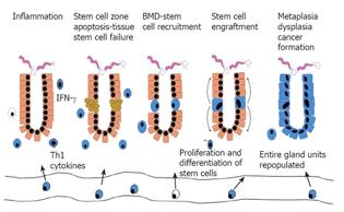
Figure 6 A new paradigm proposed for epithelial cancer.
- Citation: Li HC, Stoicov C, Rogers AB, Houghton J. Stem cells and cancer: Evidence for bone marrow stem cells in epithelial cancers. World J Gastroenterol 2006; 12(3): 363-371
- URL: https://www.wjgnet.com/1007-9327/full/v12/i3/363.htm
- DOI: https://dx.doi.org/10.3748/wjg.v12.i3.363


