Copyright
©2006 Baishideng Publishing Group Co.
World J Gastroenterol. Aug 7, 2006; 12(29): 4754-4756
Published online Aug 7, 2006. doi: 10.3748/wjg.v12.i29.4754
Published online Aug 7, 2006. doi: 10.3748/wjg.v12.i29.4754
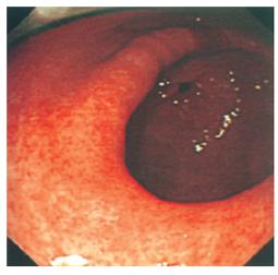
Figure 1 Gastric antrum viewed from mid body of stomach showing widespread erythematous changes consistent with gastritis.
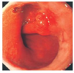
Figure 2 Gastro-oesophageal junction showing polypoid lesion.
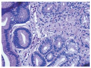
Figure 3 Gastric antral biopsy (HE x 400 magnification) showing surface epithelium on the left of the image.
The glands are displaced by a central granuloma formed from loosely aggregated epithelioid histiocytes, with a few surrounding lymphocytes.
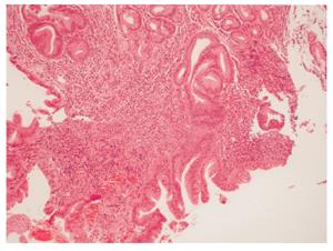
Figure 4 Biopsy of polypoid lesion at gastro-oesophageal junction showing glandular tissue with active chronic inflammation and reactive epithlelial changes (HE x 200 magnification).
- Citation: Leeds JS, McAlindon ME, Lorenz E, Dube AK, Sanders DS. Gastric sarcoidosis mimicking irritable bowel syndrome-Cause not association? World J Gastroenterol 2006; 12(29): 4754-4756
- URL: https://www.wjgnet.com/1007-9327/full/v12/i29/4754.htm
- DOI: https://dx.doi.org/10.3748/wjg.v12.i29.4754


