Copyright
©2006 Baishideng Publishing Group Co.
World J Gastroenterol. Jul 28, 2006; 12(28): 4596-4598
Published online Jul 28, 2006. doi: 10.3748/wjg.v12.i28.4596
Published online Jul 28, 2006. doi: 10.3748/wjg.v12.i28.4596
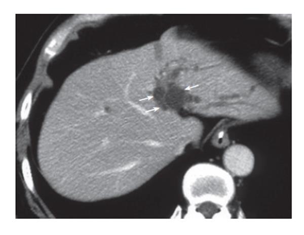
Figure 1 CT revealing multiple small cystic lesions along the umbilical portion of the portal vein (arrows).
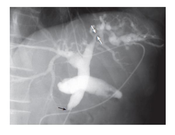
Figure 2 Cholangiogram via endoscopic naso-biliary drainage tube revealing complete obstruction in the lower bile duct (black arrow) and smooth stenosis of the left hepatic duct (white arrows) and a slight dilatation of the intrahepatic bile duct in the left lobe.
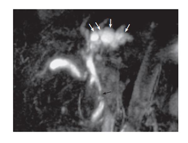
Figure 3 Magnetic resonance cholangiopancreatogram showing bead-like cystic lesions (arrows) along the biliary tree in the left lobe of the liver.
- Citation: Miura F, Takada T, Amano H, Yoshida M, Isaka T, Toyota N, Wada K, Takagi K, Kato K. A case of peribiliary cysts accompanying bile duct carcinoma. World J Gastroenterol 2006; 12(28): 4596-4598
- URL: https://www.wjgnet.com/1007-9327/full/v12/i28/4596.htm
- DOI: https://dx.doi.org/10.3748/wjg.v12.i28.4596


