Copyright
©2006 Baishideng Publishing Group Co.
World J Gastroenterol. Jul 21, 2006; 12(27): 4437-4439
Published online Jul 21, 2006. doi: 10.3748/wjg.v12.i27.4437
Published online Jul 21, 2006. doi: 10.3748/wjg.v12.i27.4437
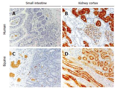
Figure 1 AQP1 expression in human and equine small intestine and kidney.
In the small intestine of both species, AQP1 is present exclusively in microvessel endothelia and in erythrocytes (panels A and C). AQP1 is not expressed in epithelial cells or any other structures in the small intestine (panels A and C). In the kidney of both species specific AQP1 expression is abundant in the renal cortex, specifically in the apical (brush border) and basolateral membranes of proximal tubules and in microvessel endothelia (panels B and D). Some AQP1 expression is also seen within the glomerulus in podocytes (panels B and D).
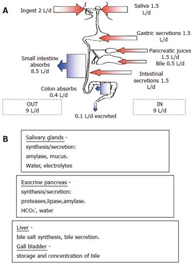
Figure 2 Summary of the gastrointestinal system’s daily fluid balance (A) and the secretions of salivary glands, exocrine pancreas and liver (B).
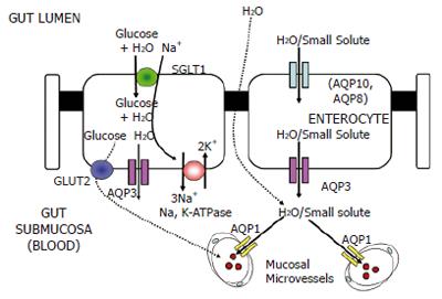
Figure 3 Model of water and small solute transport across the absorptive epithelium of the human small intestine[21].
The potential routes for water entry into the enterocyte from the luminal (apical) side are AQP10 and the sodium/glucose co-transporter SGLT1. SGLT1 is a secondary active transporter driven by the function of basolateral Na,K-ATPase which co-transports Na+ and glucose along with water into enterocytes. AQP8 (shown in parentheses) has been shown to be located in the sub-apical cytoplasm of absorptive epithelial cells. The basolateral water exit pathway is most likely to be the AQP3 protein[10,27]. Although the location of the AQP7 protein has not yet been precisely determined, it may be involved in glycerol transport in the intestinal lacteal system.
- Citation: Mobasheri A. Comment on: Cloning and characterization of porcine aquaporin 1 water channel expressed extensively in the gastrointestinal system. World J Gastroenterol 2006; 12(27): 4437-4439
- URL: https://www.wjgnet.com/1007-9327/full/v12/i27/4437.htm
- DOI: https://dx.doi.org/10.3748/wjg.v12.i27.4437


