Copyright
©2006 Baishideng Publishing Group Co.
World J Gastroenterol. Jul 21, 2006; 12(27): 4431-4434
Published online Jul 21, 2006. doi: 10.3748/wjg.v12.i27.4431
Published online Jul 21, 2006. doi: 10.3748/wjg.v12.i27.4431
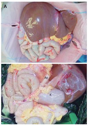
Figure 1 Graft after perfusion and removal including liver stomach, duodenal-pancreatic complex, small intestine, ascending colon and right kidney (K).
The greater omentum (O) was preserved to protect the bowels in case of abdominal dehiscence. The spleen was removed at the back-table. The aorta (Ao) was sutured below the offspring of the right renal artery and used as a neo-colic trunk.
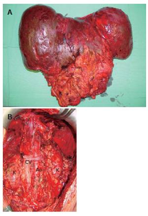
Figure 2 Recipient operation with a meticulous surgical dissection and exenteration of the abdominal cavity.
A: Recipient’s steatotic liver and reminiscence of mesenterium and duodenal-pancreatic complex; B: During exenteration caval vein (CV) was resected and prepared for the orthotopic placement of the multivisceral graft. The aorta (Ao) was preserved in full length and both kidneys (K) of the recipient were left in place.
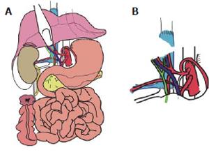
Figure 3 Venous and arterial reconstruction.
A: Schematic demonstration of the multivisceral transplant. Venous reconstruction was accomplished by inserting the retrohepatic caval vein including the right renal vein (RV) into the recipient (CV) using standard end-to-end anastomosis of the suprahepatic inferior CV and of the infrahepatic CV between the insertion of the right donor RV and recipient RV. B: Arterial reconstruction was performed using the donor abdominal aorta with the natural offsprings of the celiac trunk (CT), superior mesenteric artery (SMA), and right renal artery (RA) and inserting it as aneo-celiac trunk at the site of the original recipient celiac trunk.
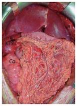
Figure 4 Situs after reperfusion.
The right kidney (KTx) including the adrenal gland was grafted orthotopically. The greater omentum was used to cover the small bowel and the ascending colon (Co) to protect it against direct contact with the alloplastic mesh.
- Citation: Pascher A, Klupp J, Kohler S, Langrehr JM, Neuhaus P. Transplantation of an eight-organ multivisceral graft in a patient with frozen abdomen after complicated Crohn’s disease. World J Gastroenterol 2006; 12(27): 4431-4434
- URL: https://www.wjgnet.com/1007-9327/full/v12/i27/4431.htm
- DOI: https://dx.doi.org/10.3748/wjg.v12.i27.4431


