Copyright
©2006 Baishideng Publishing Group Co.
World J Gastroenterol. Jul 21, 2006; 12(27): 4425-4427
Published online Jul 21, 2006. doi: 10.3748/wjg.v12.i27.4425
Published online Jul 21, 2006. doi: 10.3748/wjg.v12.i27.4425
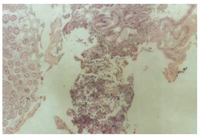
Figure 1 Primary malignant melanoma of the stomach.
Gastric mucosa also viewed (HE, × 40).
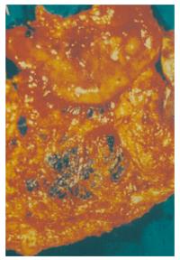
Figure 2 Surgical specimen of a subtotal gastrectomy and splenectomy for a primary melanoma located at the antrum of the stomach.
Certain deposits of malignant melanocytes are viewed in the co-excised greater omentum.
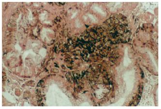
Figure 3 Primary gastric melanoma stained with Masson-Fontana.
Plenty of granules of melanin are viewed (× 100).
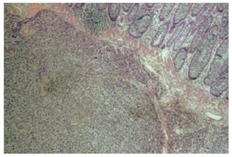
Figure 4 Primary gastric melanoma stained with HMB-45 (× 100).
- Citation: Lagoudianakis EE, Genetzakis M, Tsekouras DK, Papadima A, Kafiri G, Toutouzas K, Katergiannakis V, Manouras A. Primary gastric melanoma: A case report. World J Gastroenterol 2006; 12(27): 4425-4427
- URL: https://www.wjgnet.com/1007-9327/full/v12/i27/4425.htm
- DOI: https://dx.doi.org/10.3748/wjg.v12.i27.4425


