Copyright
©2006 Baishideng Publishing Group Co.
World J Gastroenterol. Jul 21, 2006; 12(27): 4401-4405
Published online Jul 21, 2006. doi: 10.3748/wjg.v12.i27.4401
Published online Jul 21, 2006. doi: 10.3748/wjg.v12.i27.4401
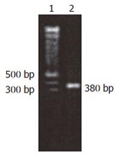
Figure 1 PCR 1.
5% Gel electro-phoresis. Lane1: 100 bp Ladder Marker; Lane2: hAL-RcDNA.
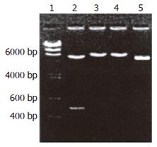
Figure 2 pGEX-4T-2-hALR digestion analysis.
Lane1: λDNA/HindIII marker; Lane2: pGEX-4T-2-hALR/B, E; Lane3: pGEX-4T-2-hALR/BamHI; Lane4: pGEX-4T-2-hALR/EcoR I; Lane5: pGEX-4T-2-/B, E. (B: BamHI, E: EcoRI).
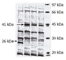
Figure 3 Expression of GST-hALR fusion protein.
Lane1: marker; Lane2: induced 1h; Lane3: induced 3h; Lane4: induced 8h; Lane5: control.
Figure 4 Comparison of inclusion body and supernatant.
Lane1: marker; Lane2: inclusion body; Lane3: supernatan.
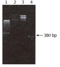
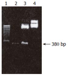
Figure 5 Digestion pEGFP-C1 and hALR.
Lane1: 100 bp Ladder Marker; Lane2: pEGFP-c1 E/B; Lane3: pEGFP-c1; Lane4: hALR E/B.
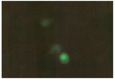
Figure 6 Expression of green fluorescence protein of recombinant pEGFP- c1-ALR in LO2 hepatocyte.
- Citation: Zhang YD, Zhou J, Zhao JF, Peng J, Liu XD, Liu XS, Jia ZM. Expression, purification and bioactivity of human augmenter of liver regeneration. World J Gastroenterol 2006; 12(27): 4401-4405
- URL: https://www.wjgnet.com/1007-9327/full/v12/i27/4401.htm
- DOI: https://dx.doi.org/10.3748/wjg.v12.i27.4401


