Copyright
©2006 Baishideng Publishing Group Co.
World J Gastroenterol. Jul 14, 2006; 12(26): 4273-4275
Published online Jul 14, 2006. doi: 10.3748/wjg.v12.i26.4273
Published online Jul 14, 2006. doi: 10.3748/wjg.v12.i26.4273
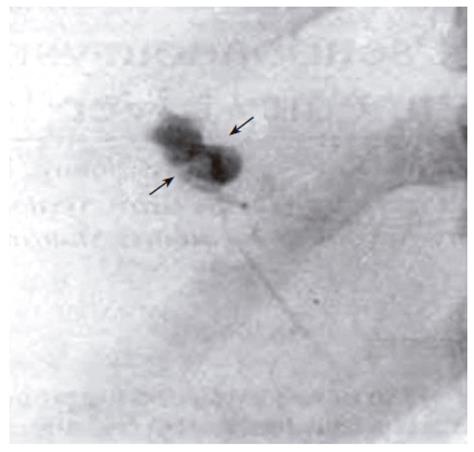
Figure 1 Angiography of the right hepatic artery.
Pseudoaneurysm (arrows) in the right hepatic artery branch.
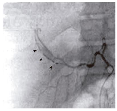
Figure 2 Platinum coils (arrows) are seen filling the feeding branch.
Angiography obtained after embolization of the branch leading to and from the pseudoaneurysm.
- Citation: Ren FY, Piao XX, Jin AL. Delayed hemorrhage from hepatic artery after ultrasound-guided percutaneous liver biopsy: A case report. World J Gastroenterol 2006; 12(26): 4273-4275
- URL: https://www.wjgnet.com/1007-9327/full/v12/i26/4273.htm
- DOI: https://dx.doi.org/10.3748/wjg.v12.i26.4273


