Copyright
©2006 Baishideng Publishing Group Co.
World J Gastroenterol. Jul 14, 2006; 12(26): 4270-4272
Published online Jul 14, 2006. doi: 10.3748/wjg.v12.i26.4270
Published online Jul 14, 2006. doi: 10.3748/wjg.v12.i26.4270
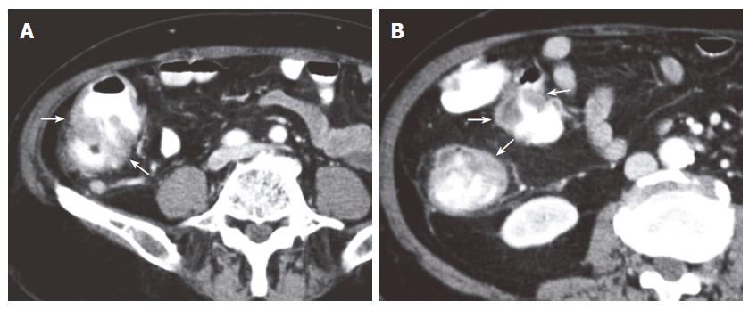
Figure 1 Computed tomographic scans (A, B) of the abdomen.
Note the irregular and nodular thickened wall of the cecum and the ascending colon (arrows).
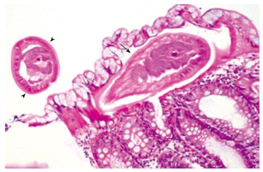
Figure 2 Pathologic examination reveals transverse section of anterior portion of Trichuris trichiura (arrow) embedded in superficial colonic mucosa and free body part of Trichuris trichiura (arrowheads) extending into the lumen of the colon.
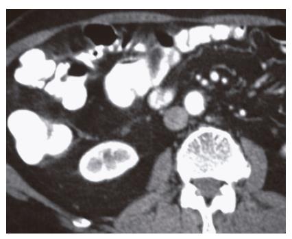
Figure 3 Computed tomographic scan obtained 6 mo after the treatment show a normal colonic wall thickness.
- Citation: Tokmak N, Koc Z, Ulusan S, Koltas IS, Bal N. Computed tomographic findings of trichuriasis. World J Gastroenterol 2006; 12(26): 4270-4272
- URL: https://www.wjgnet.com/1007-9327/full/v12/i26/4270.htm
- DOI: https://dx.doi.org/10.3748/wjg.v12.i26.4270


