Copyright
©2006 Baishideng Publishing Group Co.
World J Gastroenterol. Jul 14, 2006; 12(26): 4267-4269
Published online Jul 14, 2006. doi: 10.3748/wjg.v12.i26.4267
Published online Jul 14, 2006. doi: 10.3748/wjg.v12.i26.4267
Figure 1 Megaloblastic changes in the bone marrow.
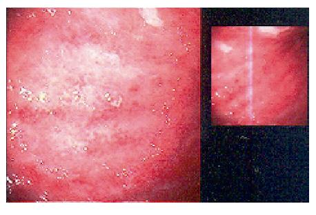
Figure 2 Polypoid appearance and atrophic gastritis at endoscopy.
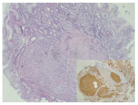
Figure 3 Carcinoid tumor positive stained with hematoxylin-eosin A and synaptophysin (x 40).
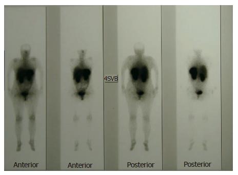
Figure 4 Indium-111 octreotide scan of the patient.
- Citation: Kadikoylu G, Yavasoglu I, Yukselen V, Ozkara E, Bolaman Z. Treatment of solitary gastric carcinoid tumor by endoscopic polypectomy in a patient with pernicious anemia. World J Gastroenterol 2006; 12(26): 4267-4269
- URL: https://www.wjgnet.com/1007-9327/full/v12/i26/4267.htm
- DOI: https://dx.doi.org/10.3748/wjg.v12.i26.4267


