Copyright
©2006 Baishideng Publishing Group Co.
World J Gastroenterol. Jul 14, 2006; 12(26): 4259-4261
Published online Jul 14, 2006. doi: 10.3748/wjg.v12.i26.4259
Published online Jul 14, 2006. doi: 10.3748/wjg.v12.i26.4259
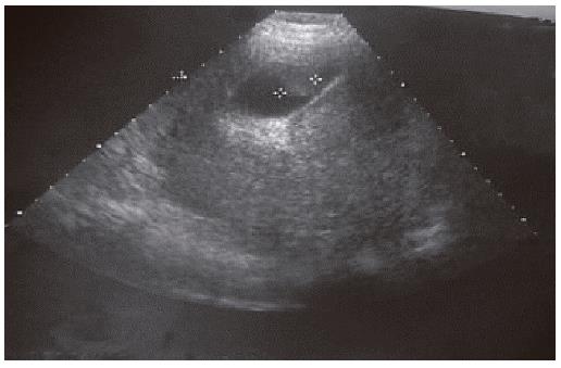
Figure 1 Ultrasound of melanoma of gallbladder.
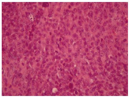
Figure 2 Histological appearance of melanoma of gallbladder stained with HE.
× 400.
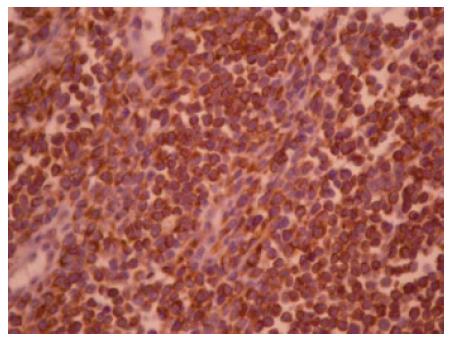
Figure 3 Neoplastic cells immunostained with anti-HMB-45 antibodies.
× 400.
- Citation: Safioleas M, Agapitos E, Kontzoglou K, Stamatakos M, Safioleas P, Mouzopoulos G, Kostakis A. Primary melanoma of the gallbladder: Does it exist? Report of a case and review of the literature. World J Gastroenterol 2006; 12(26): 4259-4261
- URL: https://www.wjgnet.com/1007-9327/full/v12/i26/4259.htm
- DOI: https://dx.doi.org/10.3748/wjg.v12.i26.4259


