Copyright
©2006 Baishideng Publishing Group Co.
World J Gastroenterol. Jul 14, 2006; 12(26): 4250-4252
Published online Jul 14, 2006. doi: 10.3748/wjg.v12.i26.4250
Published online Jul 14, 2006. doi: 10.3748/wjg.v12.i26.4250
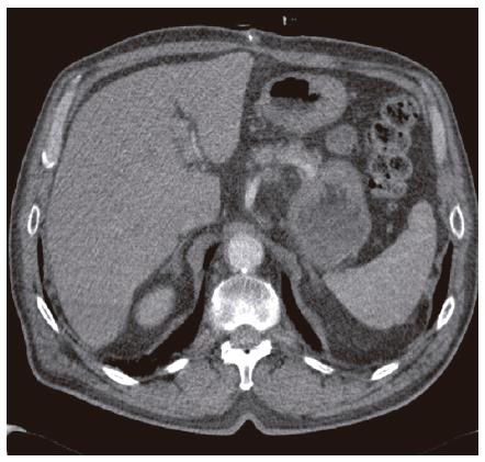
Figure 1 Preoperative CT scan (patient 1).
No parenchymal inhomogeneity or focal lesions can be detected.
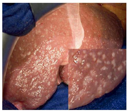
Figure 2 Intraoperative aspects (patient 1).
Multiple small nodules are present in both lobes of the liver, mimicking diffuse metastatic lesions.
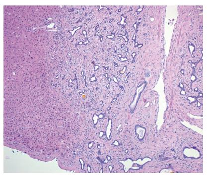
Figure 3 Histologic staining of a von Meyenburg complex (patient 1).
- Citation: Fritz S, Hackert T, Blaker H, Hartwig W, Schneider L, Buchler MW, Werner J. Multiple von Meyenburg complexes mimicking diffuse liver metastases from esophageal squamous cell carcinoma. World J Gastroenterol 2006; 12(26): 4250-4252
- URL: https://www.wjgnet.com/1007-9327/full/v12/i26/4250.htm
- DOI: https://dx.doi.org/10.3748/wjg.v12.i26.4250


