Copyright
©2006 Baishideng Publishing Group Co.
World J Gastroenterol. Jul 7, 2006; 12(25): 3994-3998
Published online Jul 7, 2006. doi: 10.3748/wjg.v12.i25.3994
Published online Jul 7, 2006. doi: 10.3748/wjg.v12.i25.3994
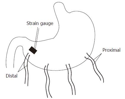
Figure 1 Surgical preparations of stomach.
Four pairs of temporary electrodes were implanted along the greater curvature at a distance of 4 cm. The most distal pair was 2 cm above the pylorus. A strain gauge was sutured on the serosal wall parallel to the most distal pair of electrodes.
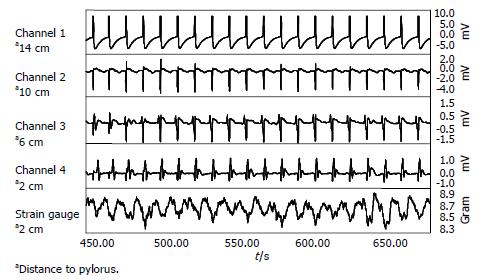
Figure 2 Sample tracings of gastric myoelectric activity recorded from implanted temporary electrodes and gastric contractions from the attached gastric strain gauge.
Top 4 channels are gastric myoelectrical activities recorded using serosal electrodes implanted along the greater curvature. The bottom tracing shows gastric contractions measured from a strain gauge implanted in the distal stomach parallel to Channel 4. It was noticed that slow waves propagate from proximal to distal stomach and each gastric slow wave was coupled with a gastric contractile event.
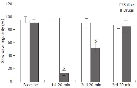
Figure 3 The regularity of gastric myoelectric activity was reduced with and after infusion of vasopressin, and returned somewhat during the last 20-min recovery period.
ANOVA, bP < 0.001 baseline vs 2nd or 3rd 20-min period (n = 7).
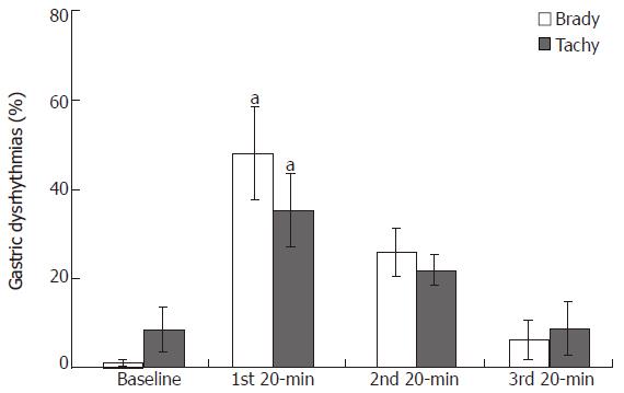
Figure 4 Distribution of gastric dysrhythmias induced by intravenous infusion of vasopressin.
Similar percentage of tachygastria and bradygastria was observed, which contributed to the irregularity of gastric myoelectric activity. ANOVA, aP < 0.05 vs baseline and 3rd 20-min period (n = 7).
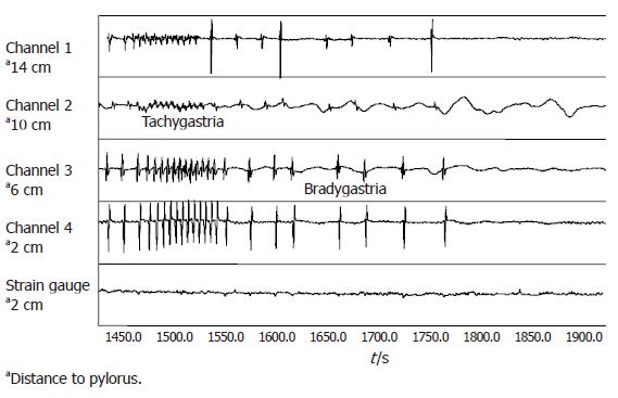
Figure 5 Illustration of gastric dysrhythmias induced by intravenous Vasopressin.
Top 4 channels are gastric myoelectrical activities recorded using serosal electrodes implanted along the greater curvature. The bottom tracing shows gastric contractions measured from a strain gauge implanted in the distal stomach parallel to Channel 4. It was noticed that gastric muscle contractions were absent during periods of tachygastria and bradygastria.
-
Citation: Xing J, Qian L, Chen J. Experimental gastric dysrhythmias and its correlation with
in vivo gastric muscle contractions. World J Gastroenterol 2006; 12(25): 3994-3998 - URL: https://www.wjgnet.com/1007-9327/full/v12/i25/3994.htm
- DOI: https://dx.doi.org/10.3748/wjg.v12.i25.3994


