Copyright
©2006 Baishideng Publishing Group Co.
World J Gastroenterol. Jul 7, 2006; 12(25): 3970-3976
Published online Jul 7, 2006. doi: 10.3748/wjg.v12.i25.3970
Published online Jul 7, 2006. doi: 10.3748/wjg.v12.i25.3970
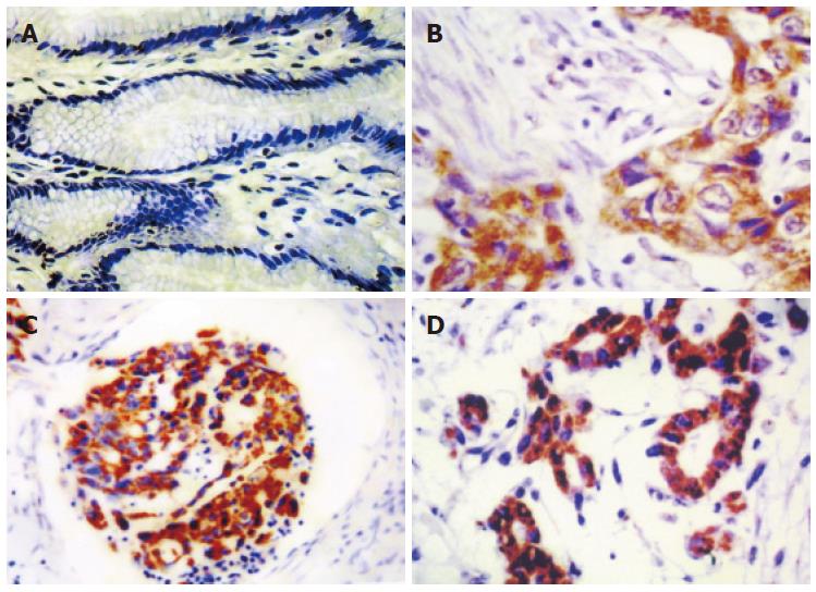
Figure 1 In situ hybridization of uPA mRNA in gastric cancer tissue.
A: Negative in the plasma of non-tumor gastric mucosa (ISH × 100); B: Positive in the plasma of gastric adenocarcinoma (ISH × 220); C: Positive in plasma of gastric adenocarcinoma with lymphangial cancer embolus (ISH × 180); D: Positive in the plasma of gastric adenocarcinoma with greater peritoneum infiltration (ISH × 180).
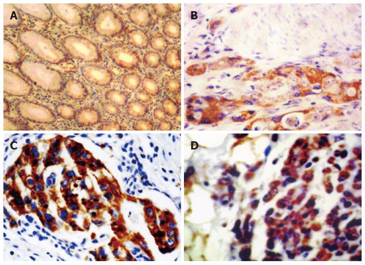
Figure 2 In situ hybridization of uPAR mRNA in gastric cancer tissue.
A: Negative in the plasma of non-tumor gastric mucosa (ISH × 100); B: Positive in the plasma of gastric adenocarcinoma infiltrated to muscularis (ISH × 180); C: Positive in plasma of gastric adenocarcinoma with lymphangial cancer embolus (ISH × 240); D: Positive in the plasma of gastric adenocarcinoma with greater peritoneum infiltration (ISH × 120).
Figure 3 VEGF expression in gastric cancer tissue.
A: Negative in the plasma of non-tumor gastric mucosa (SP×220); B: Positive in the plasma of gastric adenocarcinoma with greater omentum infiltration (SP × 220); C: Positive in plasma of gastric adenocarcinoma at the front of the cancer infiltration areas (SP × 180).
Figure 4 Microvessels in gastric cancer.
A: Negative CD34 in the vascular endothelial cell of non-tumor gastric mucosa (SP × 120); B: Positive CD34 in the vascular endothelial cell of gastric adenocarcinoma with MVD < 54.9 (SP × 400); C: Positive of CD34 in the vascular endothelial cell of gastric adenocarcinoma with MVD ≥ 54.9 (SP × 120).
Figure 5 Keplan-Meier survival curves of gastric adenocarcinoma.
A: With or without uPA mRNA (P < 0.05); B: With or without uPAR mRNA (P < 0.05); C: With or without VEGF (P < 0.05); D: With MVD ≥ 54.9 and MVD < 54.9 (P < 0.05).
- Citation: Zhang L, Zhao ZS, Ru GQ, Ma J. Correlative studies on uPA mRNA and uPAR mRNA expression with vascular endothelial growth factor, microvessel density, progression and survival time of patients with gastric cancer. World J Gastroenterol 2006; 12(25): 3970-3976
- URL: https://www.wjgnet.com/1007-9327/full/v12/i25/3970.htm
- DOI: https://dx.doi.org/10.3748/wjg.v12.i25.3970


