Copyright
©2006 Baishideng Publishing Group Co.
World J Gastroenterol. Jun 28, 2006; 12(24): 3938-3943
Published online Jun 28, 2006. doi: 10.3748/wjg.v12.i24.3938
Published online Jun 28, 2006. doi: 10.3748/wjg.v12.i24.3938
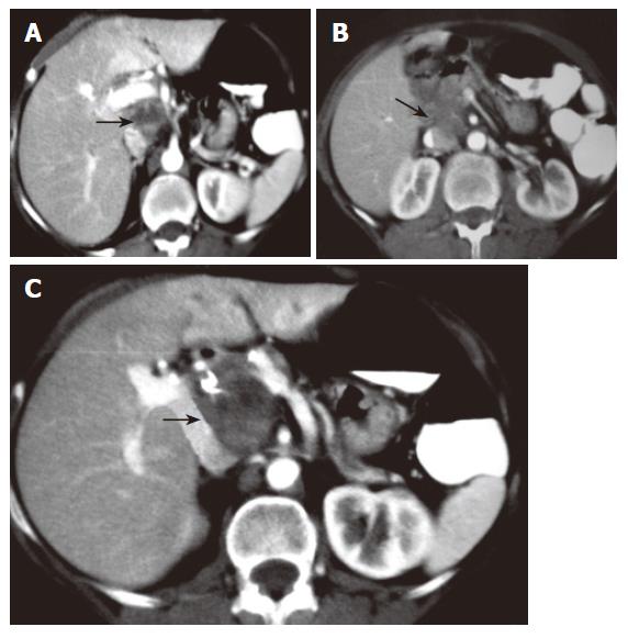
Figure 1 CT scan revealing a well delimited solid mass (arrow), without enlarged regional or para aortic lymph nodes and any evidence of invasion of other organs.
Figure 2 CT scan showing a clear relationship between tumor and inferior cava vein (arrow).
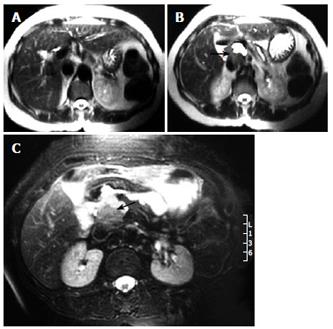
Figure 3 Serial magnetic resonance imaging of the abdomen (coronal plane) showing the tumor mass in the porta hepatis in close relation with the portal vein and biliary tract.
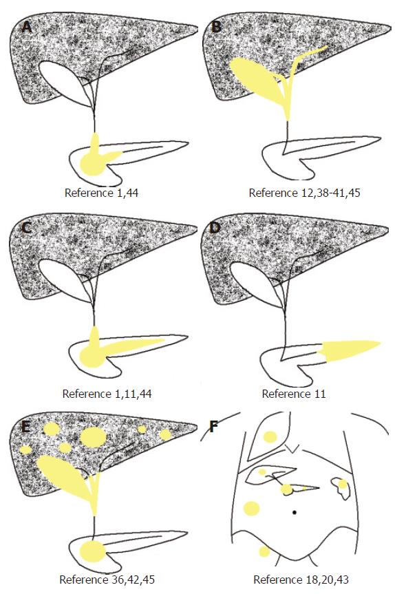
Figure 4 Dynamic classifications for autoimmune pancreatitis.
A-E: Classification of the variable involvement of the hepatobiliopancreatic system which may be observed in patients with autoimmune pancreatitis. In yellow color is shown the affected zone. Ref References of the most relevant and reliable studies that identify this specific pattern of disease. F: Multisystemic organ involvement by inflammatory pseudotumor at the biliopancreatic region. In yellow color is shown the affected zone. Ref References of the most relevant and reliable studies that identify this specific pattern of disease.
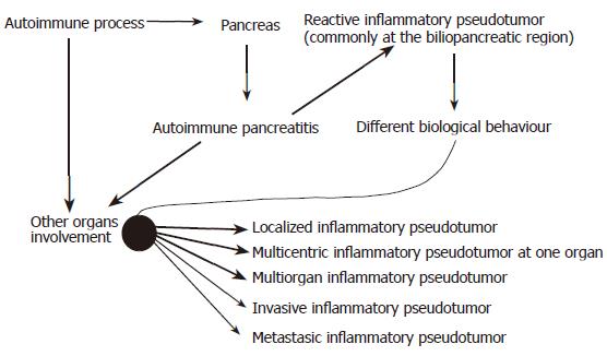
Figure 5 Autoimmune pancreatitis process.
- Citation: Fernández ELT, Luis HD, Malagón AM, González IA, Pallarés AC. Recurrence of inflammatory pseudotumor in the distal bile duct: Lessons learned from a single case and reported cases. World J Gastroenterol 2006; 12(24): 3938-3943
- URL: https://www.wjgnet.com/1007-9327/full/v12/i24/3938.htm
- DOI: https://dx.doi.org/10.3748/wjg.v12.i24.3938


