Copyright
©2006 Baishideng Publishing Group Co.
World J Gastroenterol. Jun 28, 2006; 12(24): 3835-3840
Published online Jun 28, 2006. doi: 10.3748/wjg.v12.i24.3835
Published online Jun 28, 2006. doi: 10.3748/wjg.v12.i24.3835
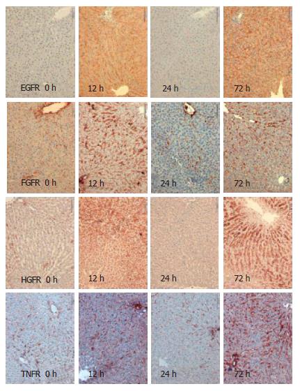
Figure 1 Sample immunohistochemistry of EGFR, FGFR, HGFR and TNFR in regenerating liver 12 h, 24 h, and 72 h after partial hepatectomy.
(0 h: control).
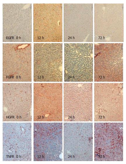
Figure 2 Sample immunohistochemistry of EGFR, FGFR, HGFR and TNFR in regenerating liver 12 h, 24 h, and 72 h after ischemia and reperfusion.
(0 h: control).
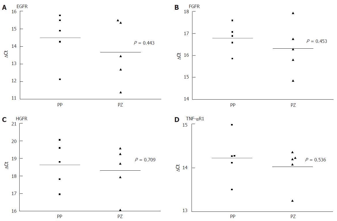
Figure 3 Results of quantitative PCR comparing receptor expression in periportal (PP) and pericentral (PC) hepatocytes in resting liver.
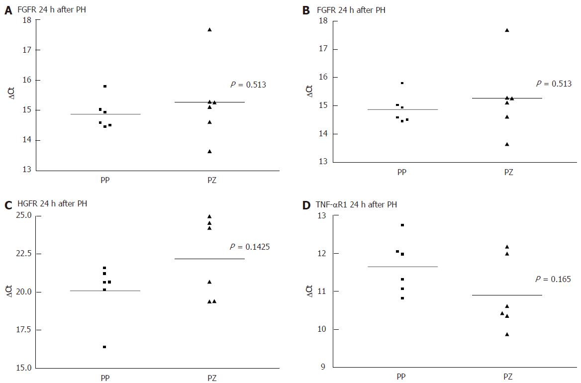
Figure 4 Results of quantitative PCR comparing receptor expression in periportal (PP)and pericentral (PC) hepatocytes 24 h after liver injury.
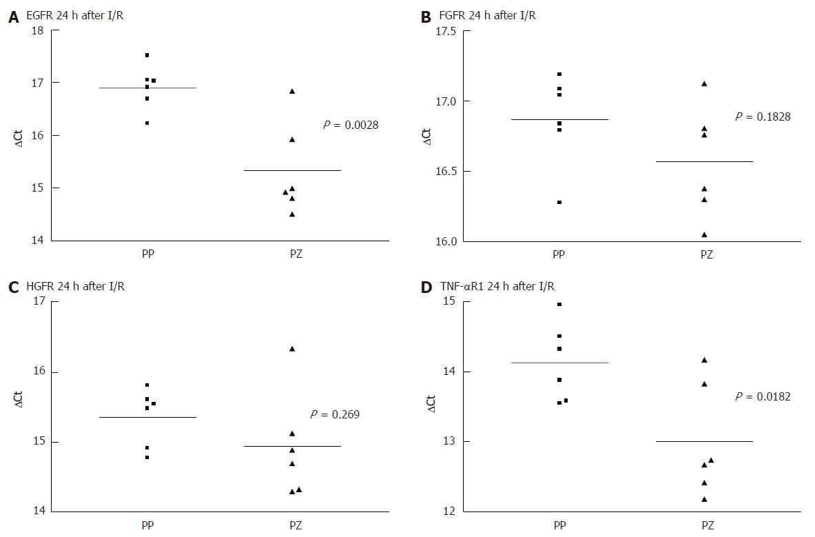
Figure 5 Results of quantitative PCR comparing receptor expression in periportal and pericentral hepatocytes 24 h after reperfusion.
- Citation: Baier P, Wolf-Vorbeck G, Hempel S, Hopt U, Dobschuetz EV. Effect of liver regeneration after partial hepatectomy and ischemia-reperfusion on expression of growth factor receptors. World J Gastroenterol 2006; 12(24): 3835-3840
- URL: https://www.wjgnet.com/1007-9327/full/v12/i24/3835.htm
- DOI: https://dx.doi.org/10.3748/wjg.v12.i24.3835


