Copyright
©2006 Baishideng Publishing Group Co.
World J Gastroenterol. Jun 21, 2006; 12(23): 3770-3775
Published online Jun 21, 2006. doi: 10.3748/wjg.v12.i23.3770
Published online Jun 21, 2006. doi: 10.3748/wjg.v12.i23.3770
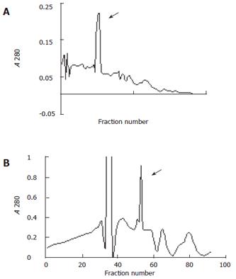
Figure 1 Elution of fractionations on cation exchange.
A: Chromatography of the ultrafiltration fraction of 35% to 50% (NH4)2SO4 ammonium sulfate precipitation (22 mg of protein ) on CM-cellulose. The procedure was performed with a column (2.6 cm × 30 cm) at a flow rate of 65 to 75 mL per hour. About 5 mL fractions was pooled. The column was developed initially with 750mL of 0.011 M citric acid-NaOH (pH 4.5), 0.02% NaN3, and then a stepwise dilution over 400 mL from 0.05, 0.10, 0.15 to 0.25 mol/L NaCl was used to accomplish the development; B: Chromatography of the ultrafiltration fraction of 50% to 60% (NH4)2SO4 ammonium sulfate precipitation (5.6 mg of protein ) on CM-cellulose. The arrows indiente the elution peak of the interest protein.
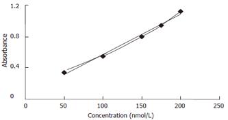
Figure 2 Stand curve of AFU.
Solid line represents an artifact line generated by the Microsoft excel™ and dot line represents the practical values in linear range.
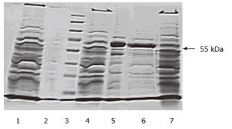
Figure 3 Electrophoretic analysis of AFU in crude and purified human liver.
Lane 1: 50% to 60% (NH4)2SO4 precipitation; lane 2: supernatant after high speed centrifugation; lane3: protein standards(Fermentas , prestained); lane 4: 35% to 50% (NH4)2SO4 precipitation; lane 5: ultrafiltration fraction after 35% to 50% (NH4)2SO4 precipitation; lane6: CM-cellulose fraction; lane 7: Homogenate and incubate at 37°C.
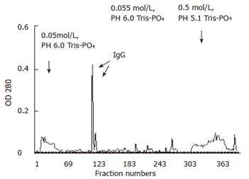
Figure 4 Anion exchange chromatography of crude antiserum against human PHCα-L-fucosidase using a column (2.
6 cm × 30 cm) of DEAE-cellulose. The column was eluted as described in Materials and methods. The arrows indicate the applied buffers and the emerged elution peak of IgG.
Figure 5 Western blot analysis showing that purified AFU could recognize a single subunit of 5 Ku (transverse arrow of the enzyme at 30s, 2 min, 5 min, respectively.
Lane 1: purified AFU (5 μg) detected with a 103 dilution of antiserum; lane 2: purified AFU (10 μg) detected with a 5×10² dilution of antiserum; lane 3: purified AFU (5 μg) detected with a 5×10² dilution of antiserum.
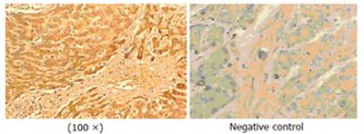
Figure 6 AFU expression in cytoplasm and/or cell membrane of PHC.
- Citation: Li C, Qian J, Lin JS. Purification and characterization of α-L-fucosidase from human primary hepatocarcinoma tissue. World J Gastroenterol 2006; 12(23): 3770-3775
- URL: https://www.wjgnet.com/1007-9327/full/v12/i23/3770.htm
- DOI: https://dx.doi.org/10.3748/wjg.v12.i23.3770


