Copyright
©2006 Baishideng Publishing Group Co.
World J Gastroenterol. Jun 14, 2006; 12(22): 3602-3608
Published online Jun 14, 2006. doi: 10.3748/wjg.v12.i22.3602
Published online Jun 14, 2006. doi: 10.3748/wjg.v12.i22.3602
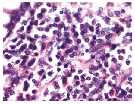
Figure 1 Photomicrograph of mucosal biopsy showing sheets of plasma cells infiltrating the lamina propria.
Epithelial cell lining the glands are well preserved (HE, × 240).
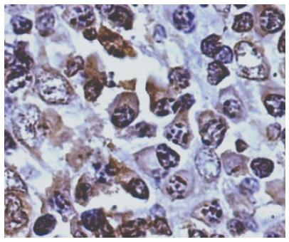
Figure 2 Photomicrograph showing strong syndecan-1-positive cells (PAP, × 440).
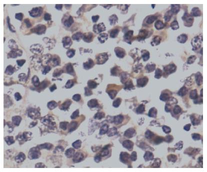
Figure 3 Photomicrograph showing bcl6-positive cells (PAP, × 440).
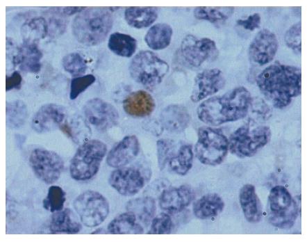
Figure 4 Photomicrograph showing p53 positivity in nuclei (PAP, × 1000).
- Citation: Vaiphei K, Kumari N, Sinha SK, Dutta U, Nagi B, Joshi K, Singh K. Roles of syndecan-1, bcl6 and p53 in diagnosis and prognostication of immunoproliferative small intestinal disease. World J Gastroenterol 2006; 12(22): 3602-3608
- URL: https://www.wjgnet.com/1007-9327/full/v12/i22/3602.htm
- DOI: https://dx.doi.org/10.3748/wjg.v12.i22.3602


