Copyright
©2006 Baishideng Publishing Group Co.
World J Gastroenterol. May 21, 2006; 12(19): 3082-3087
Published online May 21, 2006. doi: 10.3748/wjg.v12.i19.3082
Published online May 21, 2006. doi: 10.3748/wjg.v12.i19.3082
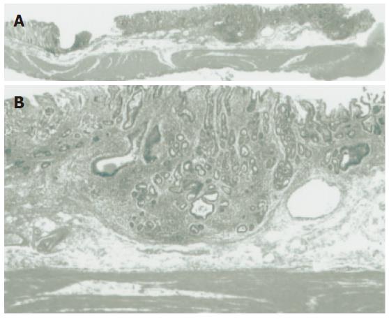
Figure 2 Histological findings.
A: Low-power histological view of a cut section of the resected specimen confirmed Dukes’ A (T1 stage) carcinoma with invasion to the submucosa. B: High-power histological view confirmed a well differentiated adenocarcinoma invading 1000 μm below the muscularis mucosa.
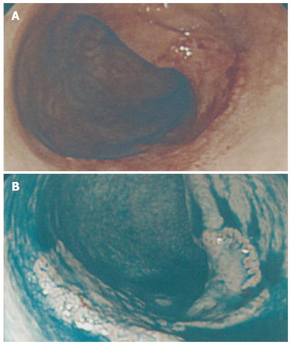
Figure 1 Superficial depressed lesion (0-IIc).
A: A superficial depressed lesion (0-IIc) of the transverse colon, 27 mm in diameter. Initially it was recognized as a broad patch of erythema and deformity of the haustrum. The patient demonstrated a family history of CRC. His mother had a past history of sigmoid colon cancer at age of 65 years. B: After dye spraying, an absolutely depressed area could be clearly identified, with a slightly elevated margin.
- Citation: Iwasaki J, Sano Y, Fu KI, Machida A, Okuno T, Kuwamura H, Yoshino T, Mera K, Kato S, Ohtsu A, Yoshida S, Fujii T. Depressed-type (0-IIc) colorectal neoplasm in patients with family history of first-degree relatives with colorectal cancer: A cross-sectional study. World J Gastroenterol 2006; 12(19): 3082-3087
- URL: https://www.wjgnet.com/1007-9327/full/v12/i19/3082.htm
- DOI: https://dx.doi.org/10.3748/wjg.v12.i19.3082


