Copyright
©2006 Baishideng Publishing Group Co.
World J Gastroenterol. May 21, 2006; 12(19): 3055-3059
Published online May 21, 2006. doi: 10.3748/wjg.v12.i19.3055
Published online May 21, 2006. doi: 10.3748/wjg.v12.i19.3055
Figure 1 Effects of rhIL-11 treatment on apoptosis and necrosis rates of IEC-6 cells 6 h after 4.
0Gy neutron irradiation. A: The apoptosis and necrosis rates of normal IEC-6 cells were 0.79% and 1.99%, respectively; B: In 4.0Gy neutron-irradiated IEC-6 cells, the apoptosis and necrosis rates were 5.58% and 9.04%, respectively; C: In IEC-6 cells treated with rhIL-11 before neutron irradiation, the apoptosis rate was 1.91%, lower than that without rhIL-11 stimulation; D: In IL-11 post-treatment IEC-6 cells, the apoptosis rate was 1.86%, lower than that without rhIL-11 stimulation. But in both rhIL-11-treated IEC-6 cells, the necrosis rate was not decreased obviously.
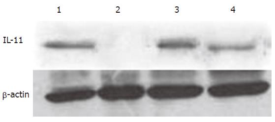
Figure 2 Effect of 4.
0Gy neutron irradiation on IL-11 expression of IEC-6 cells. Lane 1: normal control group; lanes 2-4: 6, 24 and 48 h after 4.0Gy neutron irradiation, respectively.
Figure 3 Immunohistochemichal staining of IL-11Rα (400×) located in cytomembrane and cytoplasm of IEC-6 cells.
A: IL-11Rα was strongly positive in normal IEC-6 cells; B: Six hours after 4.0Gy neutron irradiation, dramatic decline was found in IL-11Rα expression; C: No obvious decline was observed in IEC-6 cells treated with 100 ng/mL rhIL-11 after 4.0Gy neutron irradiation.
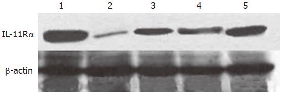
Figure 4 IL-11Rα and β-actin expressions in IEC-6 cells by Western blot.
Lane 1: normal control group; lanes 2-3: 6 and 24 h after 4.0Gy neutron irradiation; lanes 4-5: 6 and 24 h of rhIL-11 treatment after irradiation.
Figure 5 gp130 protein and β-actin expressions in IEC-6 cells by Western blot.
Lane 1: normal control group; lanes 2–4: 6, 24 and 48 h after 4.0Gy neutron irradiation respectively; lanes 5-7: 6, 24 and 48 h of rhIL-11 pretreatment group; lanes 8-10: 6, 24 and 48 h of rhIL-11 treatment after irradiation.
- Citation: Wang RJ, Peng RY, Fu KF, Gao YB, Han RG, Hu WH, Luo QL, Ma JJ. Effect of recombinant human interleukin-11 on expressions of interleukin-11 receptor α-chain and glycoprotein 130 in intestinal epithelium cell line-6 after neutron irradiation. World J Gastroenterol 2006; 12(19): 3055-3059
- URL: https://www.wjgnet.com/1007-9327/full/v12/i19/3055.htm
- DOI: https://dx.doi.org/10.3748/wjg.v12.i19.3055


