Copyright
©2006 Baishideng Publishing Group Co.
World J Gastroenterol. May 14, 2006; 12(18): 2958-2961
Published online May 14, 2006. doi: 10.3748/wjg.v12.i18.2958
Published online May 14, 2006. doi: 10.3748/wjg.v12.i18.2958
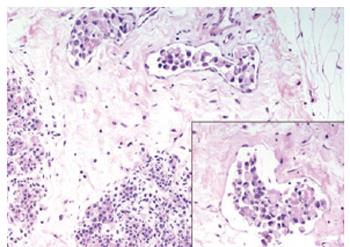
Figure 1 Breast biopsy showing breast lymphatic invasion from neoplastic cells with signet-ring features (Hematoxylin & eosin, 200 ×, 400 ×).
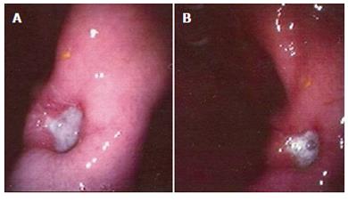
Figure 2 (A & B) Upper GI endoscopy of the patient, showing a large ulcerative lesion in the prepyloric region.
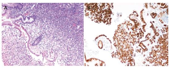
Figure 3 Gastric biopsy showing infiltration from neoplastic cells with signet-ring features (Hematoxylin & eosin, 100 ×).
Right picture shows immunohistochemical stain for CK-7 (Hematoxylin & eosin, 100 ×).
- Citation: Boutis AL, Andreadis C, Patakiouta F, Mouratidou D. Gastric signet-ring adenocarcinoma presenting with breast metastasis. World J Gastroenterol 2006; 12(18): 2958-2961
- URL: https://www.wjgnet.com/1007-9327/full/v12/i18/2958.htm
- DOI: https://dx.doi.org/10.3748/wjg.v12.i18.2958


