Copyright
©2006 Baishideng Publishing Group Co.
World J Gastroenterol. May 14, 2006; 12(18): 2825-2829
Published online May 14, 2006. doi: 10.3748/wjg.v12.i18.2825
Published online May 14, 2006. doi: 10.3748/wjg.v12.i18.2825
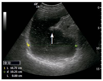
Figure 1 Ultrasonogram showing an oblique frontal section of the proximal stomach in a patient with dyspepsia referred for Ultrasound Meal Accommodation Test (UMAT).
The transversal diameter is measured indicating the size of the postprandial proximal stomach and hence indirectly degree of accommodation. Interestingly, one can also observe a gastric contraction (white arrow) in the posterior angular part and progressing towards the antrum.
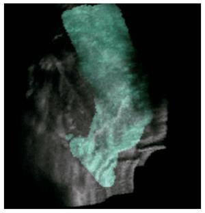
Figure 2 3D image reconstruction based on ultrasound acquisition with a magneto-based position- and orientation measurement system (POM) using GE Logiq 9.
The 3D image is overlayed the ordinary gray scale values of ultrasonograms.
- Citation: Gilja OH, Lunding J, Hausken T, Gregersen H. Gastric accommodation assessed by ultrasonography. World J Gastroenterol 2006; 12(18): 2825-2829
- URL: https://www.wjgnet.com/1007-9327/full/v12/i18/2825.htm
- DOI: https://dx.doi.org/10.3748/wjg.v12.i18.2825


