Copyright
©2006 Baishideng Publishing Group Co.
World J Gastroenterol. May 7, 2006; 12(17): 2798-2799
Published online May 7, 2006. doi: 10.3748/wjg.v12.i17.2798
Published online May 7, 2006. doi: 10.3748/wjg.v12.i17.2798
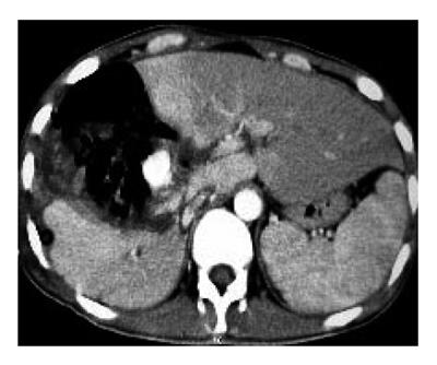
Figure 1 Axial contrast-enhanced CT scan revealing a vascular mass lesion in the right lobe of liver.
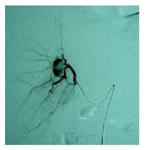
Figure 2 Selective celiac arteriogram showing a hepatic artery pseudo-aneurysm in the right hepatic artery.
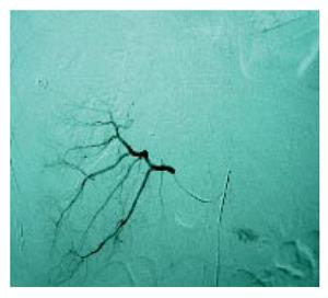
Figure 3 Post embolization selective arteriogram showing disappearance of the right hepatic artery pseudoaneurysm in the right lobe of liver.
- Citation: Sun L, Guan YS, Wu H, Pan WM, Li X, He Q, Liu Y. Post-traumatic hepatic artery pseudo-aneurysm combined with subphrenic liver abscess treated with embolization. World J Gastroenterol 2006; 12(17): 2798-2799
- URL: https://www.wjgnet.com/1007-9327/full/v12/i17/2798.htm
- DOI: https://dx.doi.org/10.3748/wjg.v12.i17.2798


