Copyright
©2006 Baishideng Publishing Group Co.
World J Gastroenterol. Apr 14, 2006; 12(14): 2301-2304
Published online Apr 14, 2006. doi: 10.3748/wjg.v12.i14.2301
Published online Apr 14, 2006. doi: 10.3748/wjg.v12.i14.2301
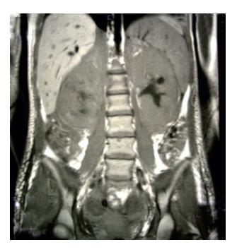
Figure 1 Magnetic resonance imaging shows extremely augmented kidneys.
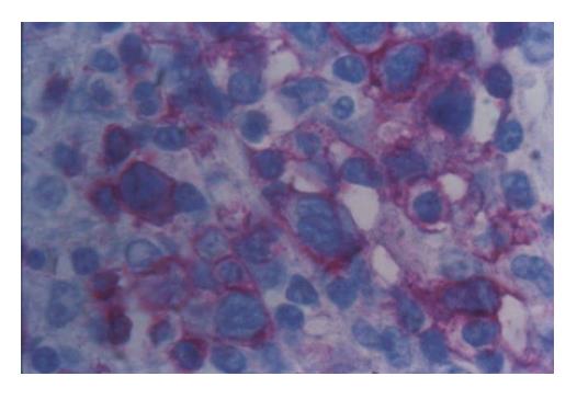
Figure 2 Neoplastic mesenterial lymph node infiltration with staining of lymphoma cells (APAAP with anti-CD3 Ab x 400).
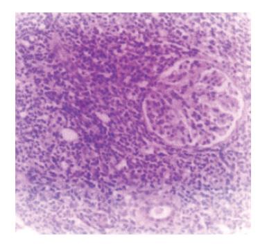
Figure 3 Renal infiltration with monomorphous lymphoid infiltration (H&E, x 40).
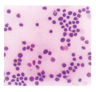
Figure 4 Neoplastic lymphoid cells in ascites (MGG, x100).
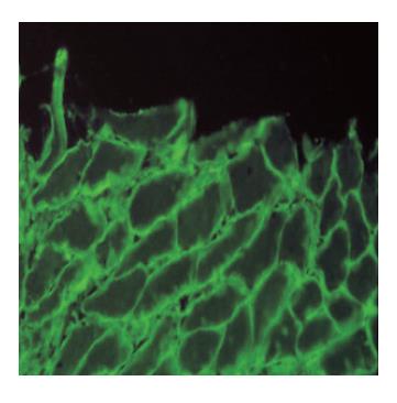
Figure 5 IgA antiendomysial antibodies surrounding sarcolemma of smoth muscle fibers in lamina muscularis mucosae of monkey esophagus (IIF, x 400).
- Citation: Bakrac M, Bonaci B, Krstic M, Simic S, Colovic M. A rare case of enteropathy-associated T-cell lymphoma presenting as acute renal failure. World J Gastroenterol 2006; 12(14): 2301-2304
- URL: https://www.wjgnet.com/1007-9327/full/v12/i14/2301.htm
- DOI: https://dx.doi.org/10.3748/wjg.v12.i14.2301


