Copyright
©2006 Baishideng Publishing Group Co.
World J Gastroenterol. Apr 14, 2006; 12(14): 2205-2208
Published online Apr 14, 2006. doi: 10.3748/wjg.v12.i14.2205
Published online Apr 14, 2006. doi: 10.3748/wjg.v12.i14.2205
Figure 1 Cystadenoma at conventional and echo-enhanced ultrasound.
A: A tumour at the pancreatic tail (5 cm in diameter) with small cystic areas (small arrows) and thin fibrotic strands; B: Highly vascularized tumour arteries (large arrows) along the fibrotic strands (maximum of contrastation 15 s after injection of the echo-enhancer).
Figure 2 Cystadenoma at conventional and echo-enhanced ultrasound A: A tumour (7 cm in diameter) at the pancreatic tail with large cystic (c) and solid areas (s); B: A poorly vascularized solid (s) tumour (maximum of contrastation 15 s after injection of the echo-enhancer).
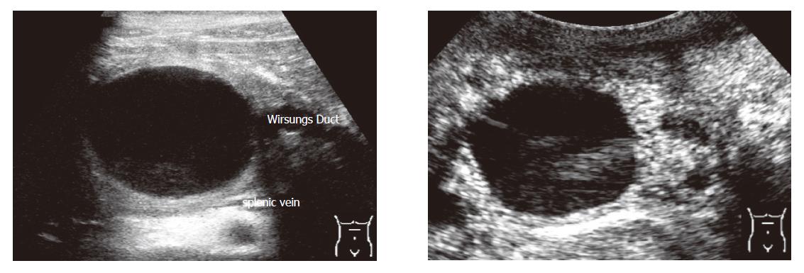
Figure 3 Pseudocust at conventional and echo-enhanced ultrasound A: A lesion with an echo-free pattern and a sharply delineated wall.
The Wirsungs Duct is dilated; B: A highly vascularized wall (maximum of contrastation 20 s after injection of the echo-enhancer).
- Citation: Rickes S, Mönkemüller K, Malfertheiner P. Echo-enhanced ultrasound with pulse inversion imaging: A new imaging modality for the differentiation of cystic pancreatic tumours. World J Gastroenterol 2006; 12(14): 2205-2208
- URL: https://www.wjgnet.com/1007-9327/full/v12/i14/2205.htm
- DOI: https://dx.doi.org/10.3748/wjg.v12.i14.2205


