Copyright
©2006 Baishideng Publishing Group Co.
World J Gastroenterol. Mar 28, 2006; 12(12): 1972-1974
Published online Mar 28, 2006. doi: 10.3748/wjg.v12.i12.1972
Published online Mar 28, 2006. doi: 10.3748/wjg.v12.i12.1972
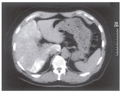
Figure 1 Non-contrast CT one month after arterial chemoembolization with iodized oil (Lipiodol) and doxorubicine emulsion.
CT shows a 2-cm nodule with dense Lipiodol enhancement in segment VI (arrow), consistent with hepatocellular carcinoma.
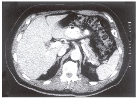
Figure 2 Follow-up contrast-enhanced CT five years later from 1995 shows a small hypervascular lesion (arrow) in segment VI consistent with residual tumor.
Tumoral Lipiodol retention was absent and the size of the lesion was significantly decreased.
- Citation: Casanovas-Taltavull T, Ribes J, Berrozpe A, Jordan S, Casanova A, Sancho C, Valls C, Bosch FX. Patient with hepatocellular carcinoma related to prior acute arsenic intoxication and occult HBV: Epidemiological, clinical and therapeutic results after 14 years of follow-up. World J Gastroenterol 2006; 12(12): 1972-1974
- URL: https://www.wjgnet.com/1007-9327/full/v12/i12/1972.htm
- DOI: https://dx.doi.org/10.3748/wjg.v12.i12.1972


