Copyright
©2006 Baishideng Publishing Group Co.
World J Gastroenterol. Mar 28, 2006; 12(12): 1969-1971
Published online Mar 28, 2006. doi: 10.3748/wjg.v12.i12.1969
Published online Mar 28, 2006. doi: 10.3748/wjg.v12.i12.1969
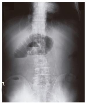
Figure 1 Plain radiography of abdomen showed stepladder appearance.
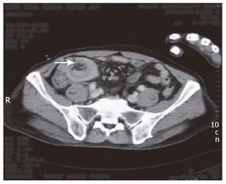
Figure 2 Computed tomography scan showed a “target mass” lesion (arrow) in the right lower abdomen, representing the intussuscepted mesenteric fat and vessel.
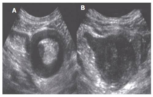
Figure 3 Abdominal ultrasound showed, A: “pseudo kidney sign” at the body of intussusception, B: Lobulated mass lesion at the head of intussusception, which was suspected of leading point of intussusception.
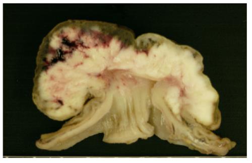
Figure 4 Surgical specimen showed that mass arising from submucosa grown into lumen and had dimpling portion on the top of tumor.
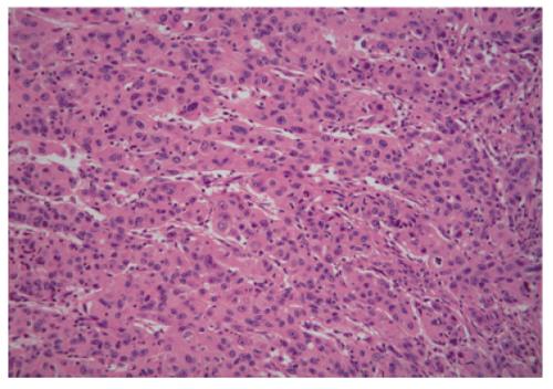
Figure 5 Histological examination demonstrated a poorly differentiated adenocarcinoma with hepatoid feature of trabecular pattern (HE X 100).
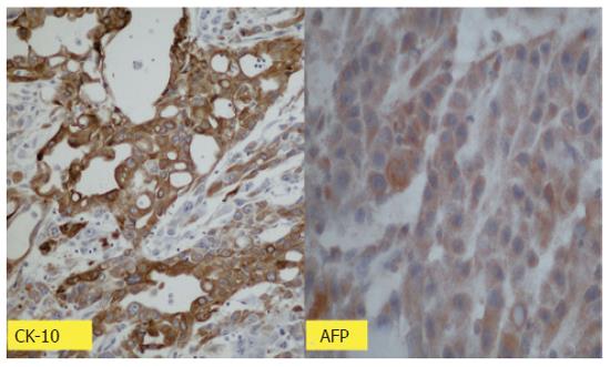
Figure 6 Strongly positive staining for alpha-fetoprotein and cytokeratin 18 (Immunoperoxidase × 100).
- Citation: Kim HS, Shin JW, Kim GY, Kim YM, Cha HJ, Jeong YK, Jeong ID, Bang SJ, Kim DH, Park NH. Metastasis of hepatocellular carcinoma to the small bowel manifested by intussusception. World J Gastroenterol 2006; 12(12): 1969-1971
- URL: https://www.wjgnet.com/1007-9327/full/v12/i12/1969.htm
- DOI: https://dx.doi.org/10.3748/wjg.v12.i12.1969


