Copyright
©2006 Baishideng Publishing Group Co.
World J Gastroenterol. Mar 28, 2006; 12(12): 1966-1968
Published online Mar 28, 2006. doi: 10.3748/wjg.v12.i12.1966
Published online Mar 28, 2006. doi: 10.3748/wjg.v12.i12.1966
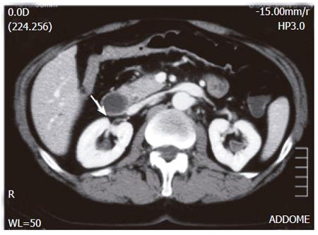
Figure 1 Evidence of polypoid mass in duodenal bulb extending to second duodenal portion (arrow).
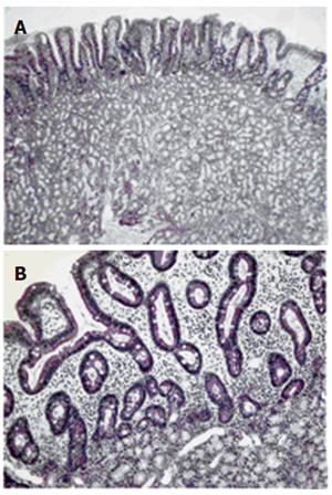
Figure 2 Gland hyperproliferation extending beyond muscolaris mucosae reaching lower portion of duodenal villi with irregular and squat profile of these structures (A) (H&E, original magnification X4) and detail of interface between Brunner’s gland proliferation and duodenal mucosa with absence of muscularis mucosae and irregular profile of villi (B) (E&E, original magnification X20).
- Citation: Rocco A, Borriello P, Compare D, Colibus PD, Pica L, Iacono A, Nardone G. Large Brunner’s gland adenoma: Case report and literature review. World J Gastroenterol 2006; 12(12): 1966-1968
- URL: https://www.wjgnet.com/1007-9327/full/v12/i12/1966.htm
- DOI: https://dx.doi.org/10.3748/wjg.v12.i12.1966


