Copyright
©2006 Baishideng Publishing Group Co.
World J Gastroenterol. Mar 21, 2006; 12(11): 1782-1785
Published online Mar 21, 2006. doi: 10.3748/wjg.v12.i11.1782
Published online Mar 21, 2006. doi: 10.3748/wjg.v12.i11.1782
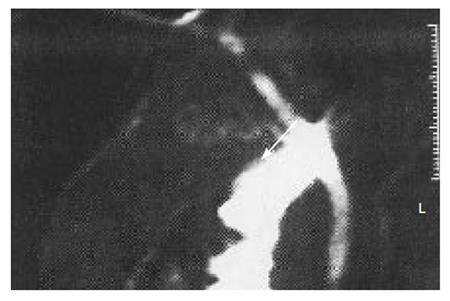
Figure 1 Cholangio-MRI showing a persistent filling defect (arrow) in the extrahepatic bile duct suspected to be biliary sludge or a stone.
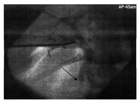
Figure 2 Intra-operative transcystic cholangiography 30 min after contrast medium injection showing the stop of contrast medium passage into the duodenum and a vanished image of the papilla of Vater (arrow) due to occluding biliary sludge.
- Citation: Greca GL, Blasi MD, Barbagallo F, Stefano MD, Latteri S, Russello D. Acute biliary pancreatitis and cholecystolithiasis in a child: One time treatment with laparoendoscopic “Rendez-vous” procedure. World J Gastroenterol 2006; 12(11): 1782-1785
- URL: https://www.wjgnet.com/1007-9327/full/v12/i11/1782.htm
- DOI: https://dx.doi.org/10.3748/wjg.v12.i11.1782


