Copyright
©2006 Baishideng Publishing Group Co.
World J Gastroenterol. Jan 7, 2006; 12(1): 150-153
Published online Jan 7, 2006. doi: 10.3748/wjg.v12.i1.150
Published online Jan 7, 2006. doi: 10.3748/wjg.v12.i1.150
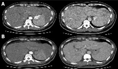
Figure 1 Multiple low-attenuating nodular lesions in the liver and an enlarged spleen (A) and their disappearance after 15 mo (B) on CT images.
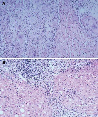
Figure 2 Broad areas of granulomatous inflammation (A) and their disappearance (B) with portal and periportal inflammation, and porto-portal fibrous septa accompanying piecemeal necrosis in hepatic lobules after 15 mo (HE ×200).
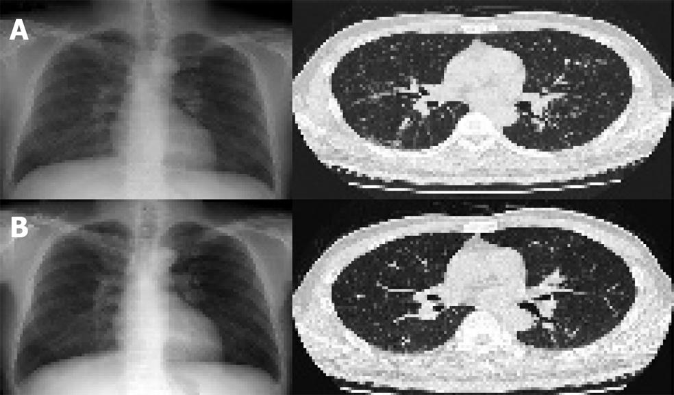
Figure 3 Scattered reticulonodular opacities and multiple fine parenchymal nodules in the case of sarcoidosis (A) and their absence after 15 mo (B).
- Citation: Kim TH, Joo JE. Spontaneous resolution of systemic sarcoidosis in a patient with chronic hepatitis C without interferon therapy. World J Gastroenterol 2006; 12(1): 150-153
- URL: https://www.wjgnet.com/1007-9327/full/v12/i1/150.htm
- DOI: https://dx.doi.org/10.3748/wjg.v12.i1.150


