Copyright
©2005 Baishideng Publishing Group Inc.
World J Gastroenterol. Mar 7, 2005; 11(9): 1403-1409
Published online Mar 7, 2005. doi: 10.3748/wjg.v11.i9.1403
Published online Mar 7, 2005. doi: 10.3748/wjg.v11.i9.1403
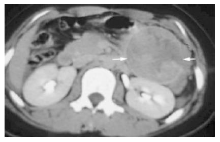
Figure 1 Computed tomographic (CT) scan of SPT.
A: Encapsulated complex, solid and cystic mass in tail of the pancreas with some internal enhancement after contrast injection shows hemorrhagic degeneration (arrow) (Case 4); B: Calcification in the mass in the body of the pancreas (arrow) (Case 6).
Figure 2 A huge complex mass with a well-defined capsule (straight arrow) and disrupted area (curved arrow) with hemoperitoneum secondary to blunt abdominal trauma (Case 2).
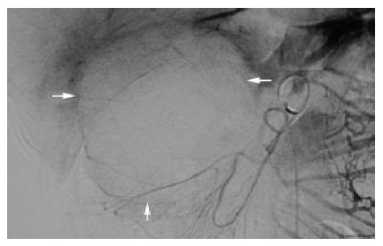
Figure 3 Superior mesenteric artery angiogram in case 3 showing avascular soft tissue lesion with displacement of nearby vessels (arrows).
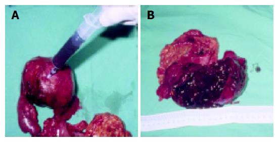
Figure 4 Gross appearance of SPT (Case 3) shows A: well encapsulated tumor with old blood clots aspirated from the lesion; B: cut section of the tumor reveals hematoma with a solid gray-white area at the periphery.
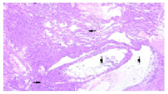
Figure 5 Tumor with pseudopapillary clusters (curved arrow), cystic spaces (straight arrow), and solid portion (arrow head) (HE, original magnification ×20).
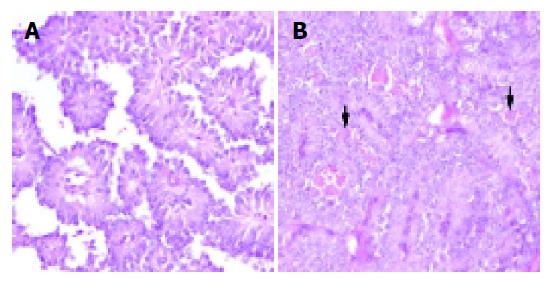
Figure 6 The tumor comprised A: branching pseudopapillary structures (HE, original magnification ×200); B: solid sheets of fairly uniform cells with cytoplasmic hyaline globules (arrows) (HE, original magnification ×200).
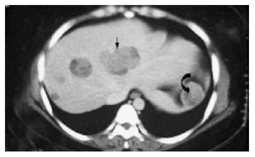
Figure 7 CT showing varying sized masses of mixed density in both lobes of the liver (straight arrow) and one similar lesion in the enlarged spleen (curved arrow) in case 1.
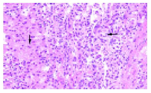
Figure 8 Metastatic tumor in liver (case 1) showing small tumor cells (straight arrow) compared to the normal liver cells with abundant cytoplasm (curved arrow) (HE, original magnification ×200).
- Citation: Huang HL, Shih SC, Chang WH, Wang TE, Chen MJ, Chan YJ. Solid-pseudopapillary tumor of the pancreas: Clinical experience and literature review. World J Gastroenterol 2005; 11(9): 1403-1409
- URL: https://www.wjgnet.com/1007-9327/full/v11/i9/1403.htm
- DOI: https://dx.doi.org/10.3748/wjg.v11.i9.1403


