Copyright
©2005 Baishideng Publishing Group Inc.
World J Gastroenterol. Feb 28, 2005; 11(8): 1096-1104
Published online Feb 28, 2005. doi: 10.3748/wjg.v11.i8.1096
Published online Feb 28, 2005. doi: 10.3748/wjg.v11.i8.1096
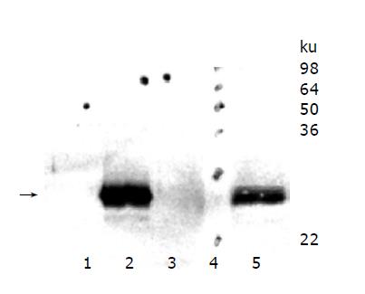
Figure 1 TIMP-1 protein expression of hepatoma cells overexpressing murine TIMP-1 and those overexpressing the TIMP-1 antagonist MMP-9-H401A.
The arrow indicates the position of murine TIMP-1. TIMP-1 protein could be detected in HepG2-TIMP-1 cells only.
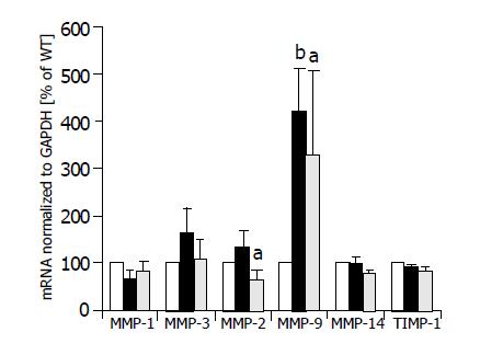
Figure 2 MMP mRNA expression of HepG2 (white columns), HepG2-TIMP-1 clones (black columns), and HepG2 cells overexpressing the TIMP-1 antagonist MMP-9-H401A (halftone columns).
aP<0.05; bP<0.01.
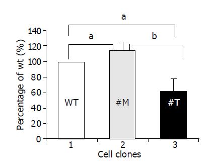
Figure 3 TIMP-1 and TIMP-1 antagonist contribute to differences in cell migration.
aP<0.05; bP<0.01.
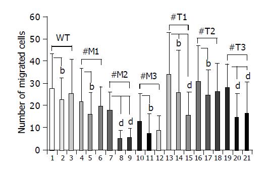
Figure 4 Effect of a broad spectrum MMP inhibitor and a specific MMP-2/MMP-9 inhibitor on the migration of HepG2, HepG2-TIMP-1 and HepG2-MMP-9-H401A cells.
bP<0.01; dP<0.001.
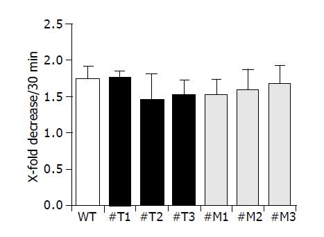
Figure 5 TIMP-1 and TIMP-1 antagonist do not influence cell attachment.
Figure 6 Effect of genistein, p38 MAPK inhibitor (SB 202190), and UO126 on the induction of MMP-2 and MMP-9 mRNA expression in HepG2 (A and C) and HepG2 TIMP-1 overexpressing cell clones (B and D).
Cells were treated with Genistein 25 µM (2), Genistein 50 µM (3), SB202190 (4), and UO126 (5). mRNA expression of untreated cells (1) were set to 100%. Incubation with DMSO served as control (6).
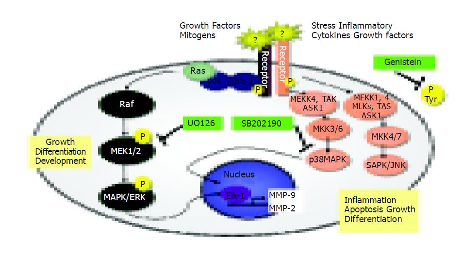
Figure 7 Signal transduction pathways and MAP kinase signaling cascades in mammalian cells shown in schematic form.
- Citation: Roeb E, Bosserhoff AK, Hamacher S, Jansen B, Dahmen J, Wagner S, Matern S. Enhanced migration of tissue inhibitor of metalloproteinase overexpressing hepatoma cells is attributed to gelatinases: Relevance to intracellular signaling pathways. World J Gastroenterol 2005; 11(8): 1096-1104
- URL: https://www.wjgnet.com/1007-9327/full/v11/i8/1096.htm
- DOI: https://dx.doi.org/10.3748/wjg.v11.i8.1096


