Copyright
©2005 Baishideng Publishing Group Inc.
World J Gastroenterol. Feb 28, 2005; 11(8): 1091-1095
Published online Feb 28, 2005. doi: 10.3748/wjg.v11.i8.1091
Published online Feb 28, 2005. doi: 10.3748/wjg.v11.i8.1091
Figure 1 A: Axial CT shows a large hyper vascularized CCC in the left liver lobe of patient 4 before therapy; B: In patient 3 axial CT shows hyper vascularized CCC nodules in the lateral right liver; C: After 7 mo of therapy there is marked shrinkage and hypodense transformation of the tumor in PR in patient 4; D: In patient 3 the same changes are apparent in the right liver in PR after 7 mo of therapy.
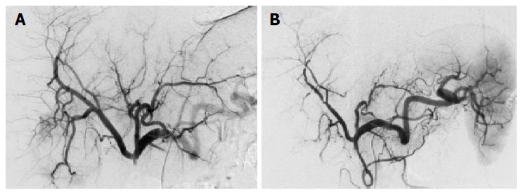
Figure 2 A: The arteriogram of patient 4 shows a beginning rarefication of the intrahepatic arteries after one chemoembolization; B: After three chemoembolizations the arteriogram of the same patient shows a progressive arterial rarefication with irregular stenoses and occlusions of peripheral branches.
- Citation: Kirchhoff T, Zender L, Merkesdal S, Frericks B, Malek N, Bleck J, Kubicka S, Baus S, Chavan A, Manns MP, Galanski M. Initial experience from a combination of systemic and regional chemotherapy in the treatment of patients with nonresectable cholangiocellular carcinoma in the liver. World J Gastroenterol 2005; 11(8): 1091-1095
- URL: https://www.wjgnet.com/1007-9327/full/v11/i8/1091.htm
- DOI: https://dx.doi.org/10.3748/wjg.v11.i8.1091


