Copyright
©2005 Baishideng Publishing Group Inc.
World J Gastroenterol. Dec 28, 2005; 11(48): 7697-7699
Published online Dec 28, 2005. doi: 10.3748/wjg.v11.i48.7697
Published online Dec 28, 2005. doi: 10.3748/wjg.v11.i48.7697
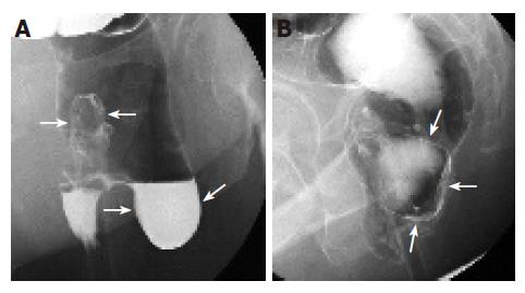
Figure 1 A and B.
Double-contrast barium enema examination demonstrates a large diverticulum with wide-orifice arising from the left lateral rectal wall (large arrows). The rectal catheter is also seen (small arrows).
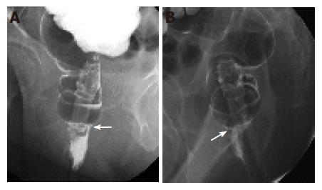
Figure 2 A and B.
Postoperative barium study revealed the subsidence of the rectal diverticulum (arrows).
- Citation: Chen CW, Jao SW, Lai HJ, Chiu YC, Kang JC. Isolated rectal diverticulum complicating with rectal prolapse and outlet obstruction: Case report. World J Gastroenterol 2005; 11(48): 7697-7699
- URL: https://www.wjgnet.com/1007-9327/full/v11/i48/7697.htm
- DOI: https://dx.doi.org/10.3748/wjg.v11.i48.7697


