Copyright
©2005 Baishideng Publishing Group Inc.
World J Gastroenterol. Dec 28, 2005; 11(48): 7684-7685
Published online Dec 28, 2005. doi: 10.3748/wjg.v11.i48.7684
Published online Dec 28, 2005. doi: 10.3748/wjg.v11.i48.7684
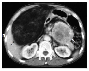
Figure 1 Abdominal CT showing the primary tumor in the body and tail of the pancreas (white arrow) and the solitary metastasis in the mesentery adjacent to the duodenum (black arrow).
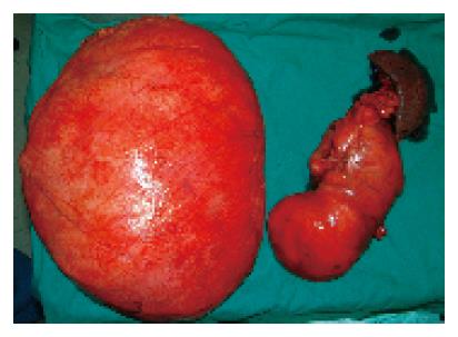
Figure 2 The primary tumor (right) and its solitary metastasis (left) after removal.
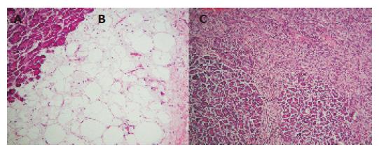
Figure 3 Microphotographs of the primary tumor.
A: Normal pancreatic tissue; B: well-differentiated sclerosing liposarcoma; C: area of dedifferentiation.
- Citation: Dodo I, Adamthwaite J, Jain P, Roy A, Guillou P, Menon K. Successful outcome following resection of a pancreatic liposarcoma with solitary metastasis. World J Gastroenterol 2005; 11(48): 7684-7685
- URL: https://www.wjgnet.com/1007-9327/full/v11/i48/7684.htm
- DOI: https://dx.doi.org/10.3748/wjg.v11.i48.7684


