Copyright
©2005 Baishideng Publishing Group Inc.
World J Gastroenterol. Dec 28, 2005; 11(48): 7625-7630
Published online Dec 28, 2005. doi: 10.3748/wjg.v11.i48.7625
Published online Dec 28, 2005. doi: 10.3748/wjg.v11.i48.7625
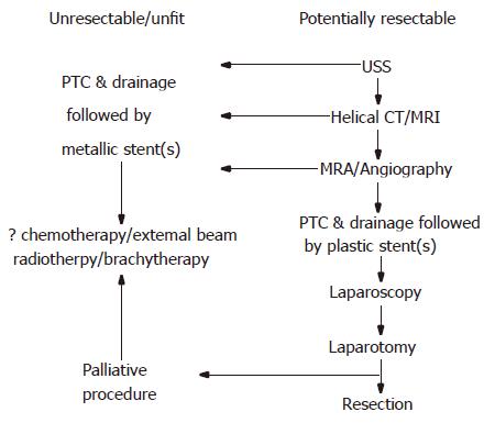
Figure 1 Flow chart demonstrating management options for hilar cholangiocarcinoma.
USS, ultrasound scan; CT, computed tomography; MRI, magnetic resonance imaging; MRA, magnetic resonance angiography; PTC, percutaneous transhepatic cholangiography.
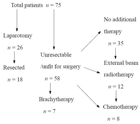
Figure 2 Flow chart showing management outcomes.
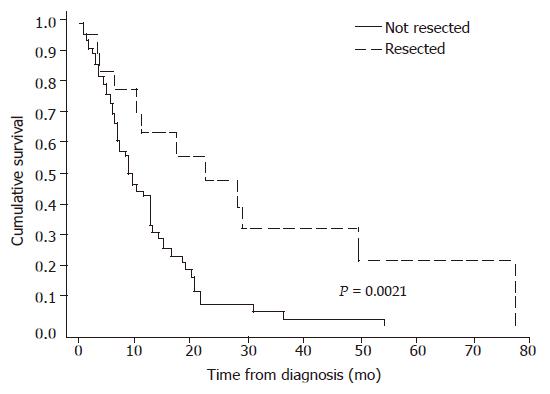
Figure 3 Kaplan–Meier survival plots for all patients with breakdown in resected and non-resected groups.
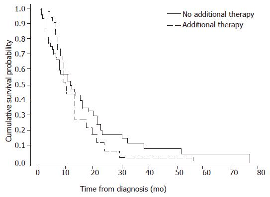
Figure 4 Kaplan-Meier survival plots for non-surgical therapeutic modalities in non-resected patients.
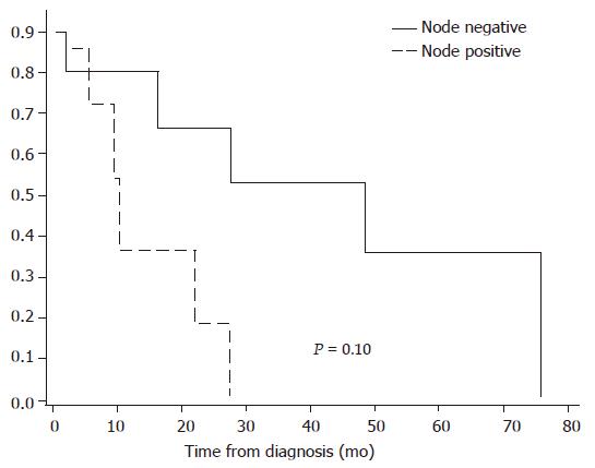
Figure 5 Kaplan-Meier survival plots for node positive and node negative resections.
- Citation: Mansfield S, Barakat O, Charnley R, Jaques B, O’Suilleabhain C, Atherton P, Manas D. Management of hilar cholangiocarcinoma in the North of England: Pathology, treatment, and outcome. World J Gastroenterol 2005; 11(48): 7625-7630
- URL: https://www.wjgnet.com/1007-9327/full/v11/i48/7625.htm
- DOI: https://dx.doi.org/10.3748/wjg.v11.i48.7625


