Copyright
©2005 Baishideng Publishing Group Inc.
World J Gastroenterol. Dec 21, 2005; 11(47): 7541-7544
Published online Dec 21, 2005. doi: 10.3748/wjg.v11.i47.7541
Published online Dec 21, 2005. doi: 10.3748/wjg.v11.i47.7541
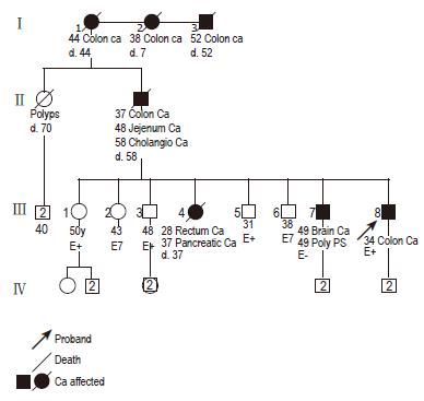
Figure 1 Pedigree of proband.
d indicates age at death. E indicates evaluation for hMSH2 mutations.
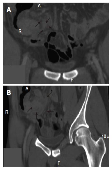
Figure 2 Spiral CT scan with multiple planar reconstruction, in coronal (2A) and oblique (2B) views, showing extrinsic localization of neoplasia.
Soft tissue density mass is visible attached to the wall of cecum (black arrows) which is compressed without clear evidence of mucosal infiltration. Air distension of cecum (A) contributes to better visualization of the mass.
- Citation: Corleto VD, Zykaj E, Mercantini P, Pilozzi E, Rossi M, Carnuccio A, Giulio ED, Ziparo V, Fave GD. Is colonoscopy sufficient for colorectal cancer surveillance in all HNPCC patients? World J Gastroenterol 2005; 11(47): 7541-7544
- URL: https://www.wjgnet.com/1007-9327/full/v11/i47/7541.htm
- DOI: https://dx.doi.org/10.3748/wjg.v11.i47.7541


