Copyright
©2005 Baishideng Publishing Group Inc.
World J Gastroenterol. Dec 14, 2005; 11(46): 7308-7313
Published online Dec 14, 2005. doi: 10.3748/wjg.v11.i46.7308
Published online Dec 14, 2005. doi: 10.3748/wjg.v11.i46.7308
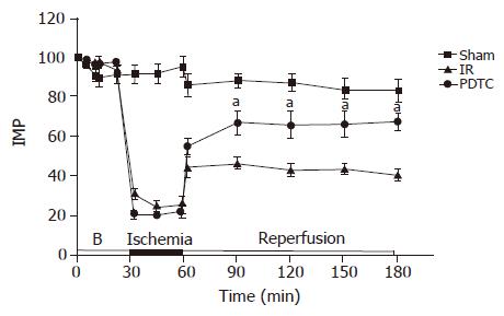
Figure 1 Intestinal microvascular perfusion in (% of baseline) during 30 min of ischemia and 2 h of reperfusion measured by L-DF.
Values are expressed as mean±SE of six animals in each group (aP<0.05 vs IR). B: Baseline.
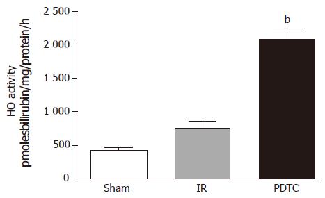
Figure 2 Ileal HO activity in all three experimental groups at the end of 2 h of reperfusion (bP<0.
01 vs IR).
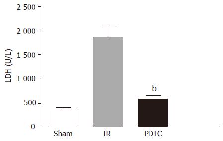
Figure 3 Serum LDH levels (U/L) at the end of 2 h reperfusion period.
Values are expressed as mean±SE of six animals in each group. In PDTC group, LDH was significantly lower compared to the IR group (bP<0.001 vs IR).
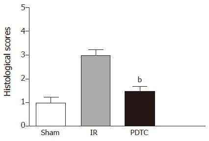
Figure 4 Comparison of histological scores of ileal mucosa between three experimental groups (bP<0.
01 vs IR).
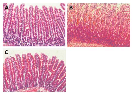
Figure 5 Representative photomicrographs of histological sections of ileum (a) in sham operated animals, (b) subjected to 30-min period of ischemia and 2-h period of reperfusion (IR) and (c) subjected to PDTC+IR (H&E, original magnification ×100).
- Citation: Mallick IH, Yang WX, Winslet MC, Seifalian AM. Pyrrolidine dithiocarbamate reduces ischemia-reperfusion injury of the small intestine. World J Gastroenterol 2005; 11(46): 7308-7313
- URL: https://www.wjgnet.com/1007-9327/full/v11/i46/7308.htm
- DOI: https://dx.doi.org/10.3748/wjg.v11.i46.7308


