Copyright
©2005 Baishideng Publishing Group Inc.
World J Gastroenterol. Dec 7, 2005; 11(45): 7084-7090
Published online Dec 7, 2005. doi: 10.3748/wjg.v11.i45.7084
Published online Dec 7, 2005. doi: 10.3748/wjg.v11.i45.7084
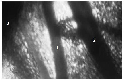
Figure 1 View of the “feeding” artery in rat’s intestinal mesentery in light microscope.
1: "feeding" vein; 2: "feeding" arteries; 3: mesenteric edge (wall) of intestine.
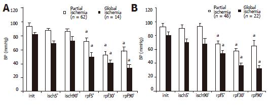
Figure 2 Systemic BPM at 90 min of partial and global intestinal ischemia and during reperfusion after 0.
9% sodium chloride injection (A) and perftoran administration (B). BPM level decreased more significantly in groups after CMA occlusion. aP<0.05 vs initial values in each group.
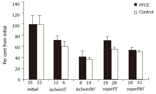
Figure 3 Diameter of “feeding” arteries at 90 min of partial intestinal ischemia and during reperfusion.
The recovery of the diameter to initial value during reperfusion was more significant in the group administered PF in comparison with control group. aP<0.05 vs control group; initial – initial values, ischem-ischemic period, reperf-reperfusion.
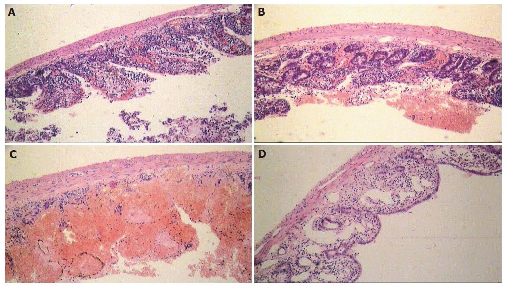
Figure 4 Changes during reperfusion following critical occlusive intestinal ischemia in rats.
A almost complete denudation and insignificant destruction of villi; B: moderate changes after 90 min of reperfusion with perftoran; C: insignificant destruction, massive hemorrhages, and focal transmural necrosis formation after 24 h of reperfusion with 0.9% sodium chloride in the intestinal wall of controls; D: initial signs of the ischemic intestinal wall regeneration in rats after 24 h of reperfusion after perftoran injection.
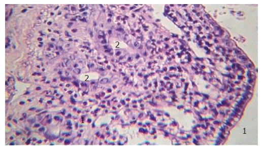
Figure 5 Villi of mucosal layer of the ischemic intestine covered by non-mature cubic epithelium after 24 h of reperfusion (1) and saved crypts in the post-ischemic period in rats administered perftoran (2).
- Citation: Kozhura VL, Basarab DA, Timkina MI, Golubev AM, Reshetnyak VI, Moroz VV. Reperfusion injury after critical intestinal ischemia and its correction with perfluorochemical emulsion "perftoran". World J Gastroenterol 2005; 11(45): 7084-7090
- URL: https://www.wjgnet.com/1007-9327/full/v11/i45/7084.htm
- DOI: https://dx.doi.org/10.3748/wjg.v11.i45.7084


