Copyright
©2005 Baishideng Publishing Group Inc.
World J Gastroenterol. Dec 7, 2005; 11(45): 7078-7083
Published online Dec 7, 2005. doi: 10.3748/wjg.v11.i45.7078
Published online Dec 7, 2005. doi: 10.3748/wjg.v11.i45.7078
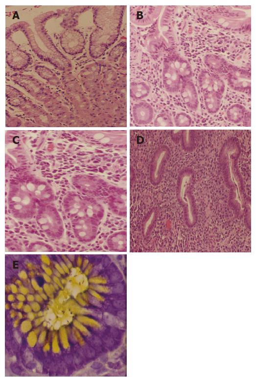
Figure 1 A: Normal histopathologic appearance of the gastric mucosa; B: Histopathology of the gastric mucosa, antral type, showing chronic inflammation with metaplasia (low power); C: Gastric mucosa, antral type (at higher power than in Figure 1B) showing chronic inflammation with intestinal metaplasia; D: Gastric biopsy showing presence of severe chronic active gastritis; E: Toluidine blue stain with alcian yellow counterstain of gastric mucosa revealing presence of H pylori.
- Citation: Dholakia K, Dharmarajan T, Yadav D, Oiseth S, Norkus E, Pitchumoni C. Vitamin B12 deficiency and gastric histopathology in older patients. World J Gastroenterol 2005; 11(45): 7078-7083
- URL: https://www.wjgnet.com/1007-9327/full/v11/i45/7078.htm
- DOI: https://dx.doi.org/10.3748/wjg.v11.i45.7078


