Copyright
©2005 Baishideng Publishing Group Inc.
World J Gastroenterol. Oct 28, 2005; 11(40): 6330-6337
Published online Oct 28, 2005. doi: 10.3748/wjg.v11.i40.6330
Published online Oct 28, 2005. doi: 10.3748/wjg.v11.i40.6330
Figure 1 General information of gene expression change.
A: Distribution of induced (black bars) and repressed genes (gray bars) based on their LSR; B: All induced and repressed genes with absolute value of LSR more than 1 after one and three doses of PG treatment and their intersection. I: One dose of PG treatment. II: Three doses of PG treatment.
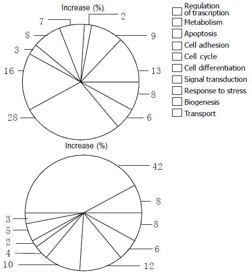
Figure 2 Functional profiles of expression changed by PG derived from Lactobacillus sp.
in peritoneal macrophages and splenic lymphocytes.
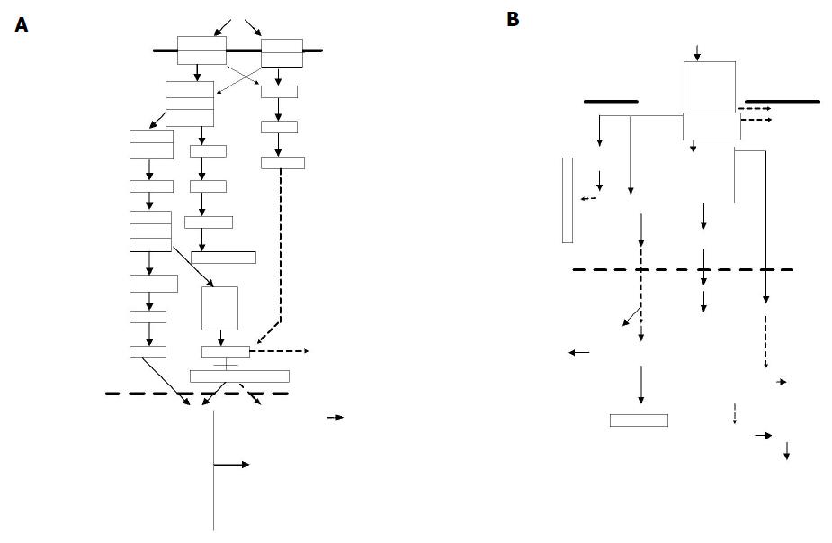
Figure 3 Functional mapping of genes to pathways for products of pro-inflammatory cytokines (A) and IFN-g and T-cell proliferation (B).
Figure 4 In vitro (A) and in vivo (B) inhibitory effects of PG on growth of CT26.
Values are mean±SD.
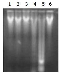
Figure 5 DNA of inoculated colon tumor in 2% agarose gel.
Lane 1-3 and 6: control; lane 4: tumor-inoculated mice treated with low doses of PG; lane 5: tumor-inoculated mice treated with high doses of PG.
-
Citation: Sun J, Shi YH, Le GW, Ma XY. Distinct immune response induced by peptidoglycan derived from
Lactobacillus sp . World J Gastroenterol 2005; 11(40): 6330-6337 - URL: https://www.wjgnet.com/1007-9327/full/v11/i40/6330.htm
- DOI: https://dx.doi.org/10.3748/wjg.v11.i40.6330


