Copyright
©The Author(s) 2005.
World J Gastroenterol. Oct 21, 2005; 11(39): 6235-6237
Published online Oct 21, 2005. doi: 10.3748/wjg.v11.i39.6235
Published online Oct 21, 2005. doi: 10.3748/wjg.v11.i39.6235
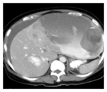
Figure 1 CT scans of the abdomen showed massive accumulation of intra-abdominal fluid and one large heterogeneous mass (c 11.
4 cm×8.8 cm×2 cm) with cystic and solid components in the left lobe of the liver.
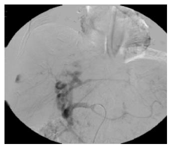
Figure 2 Catheter angiography showed a huge hepatic tumor with faint tumor blush in the left lobe and a 2.
5 cm large hypervascular tumor in the right lobe. These were presumed to be hepatocellular carcinomas.
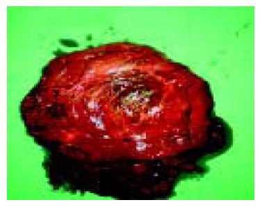
Figure 3 The specimen recovered consisted of an encapsulated mass measuring 22 cm×6 cm×5 cm in size with extensive necrosis and hemorrhage.
Grossly, it was friable and soft in consistency.
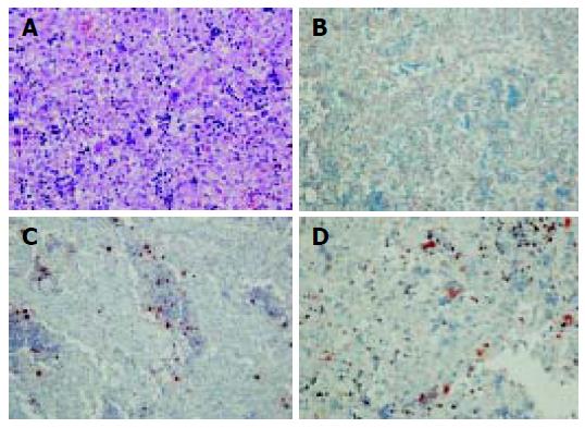
Figure 4 Immature hepatocytic epithelial elements intermixed with prominent vessels and extramedullary hematopoiesis including all three lineages (A).
Vimentin positive (B); MPO positive for the extramedullary hematopoietic myeloid series (C); factor 8 positive for extramedullary hematopoietic megakaryocytes (D).
- Citation: Ke HY, Chen JH, Jen YM, Yu JC, Hsieh CB, Chen CJ, Liu YC, Chen TW, Chan DC. Ruptured hepatoblastoma with massive internal bleeding in an adult. World J Gastroenterol 2005; 11(39): 6235-6237
- URL: https://www.wjgnet.com/1007-9327/full/v11/i39/6235.htm
- DOI: https://dx.doi.org/10.3748/wjg.v11.i39.6235


