Copyright
©The Author(s) 2005.
World J Gastroenterol. Oct 21, 2005; 11(39): 6221-6224
Published online Oct 21, 2005. doi: 10.3748/wjg.v11.i39.6221
Published online Oct 21, 2005. doi: 10.3748/wjg.v11.i39.6221
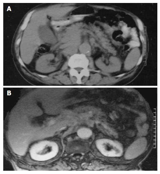
Figure 1 A: Abdominal CT scan showing the head of the pancreas and the uncinate process blurred.
B: The mass in the pancreatic head and especially the uncinate process in MRI scan.
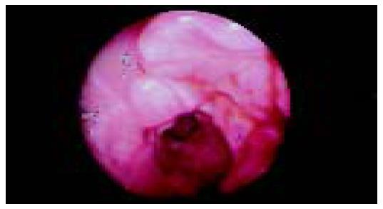
Figure 2 Endoscopic view of the ulcerated mass-like deformity of the duodenal bulb with a friable surface.
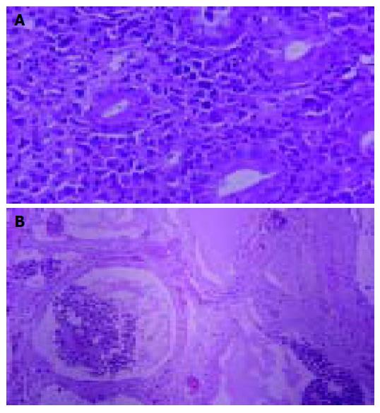
Figure 3 A: Duodenal mucosa infiltrated by polymorphous neoplastic lymphoid cells with eosinophilic cytoplasm and large nuclei.
Small numbers of acute inflammatory cells are also present (hematoxylin-eosin ×400). B: Connective tissue and vascular lumens with neoplastic emboli with similar morphologic features as in the duodenal biopsy (hematoxylin-eosin ×200).
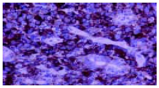
Figure 4 Strong CD30 immunohistochemical expression of lymphoma cells in the duodenal mucosa (×400).
- Citation: Savopoulos CG, Tsesmeli N, Kaiafa G, Zantidis A, Bobos M, Hatzitolios A, Papavramidis S, Kostopoulos I. Primary pancreatic anaplastic large cell lymphoma, ALK negative: A case report. World J Gastroenterol 2005; 11(39): 6221-6224
- URL: https://www.wjgnet.com/1007-9327/full/v11/i39/6221.htm
- DOI: https://dx.doi.org/10.3748/wjg.v11.i39.6221


