Copyright
©The Author(s) 2005.
World J Gastroenterol. Oct 21, 2005; 11(39): 6212-6215
Published online Oct 21, 2005. doi: 10.3748/wjg.v11.i39.6212
Published online Oct 21, 2005. doi: 10.3748/wjg.v11.i39.6212
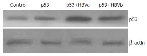
Figure 1 Western blot analysis of p53 expression in each group.
Cell lysates (80 mg) were electrophoresed on SDS-PAGE gels and proteins were transferred to PVDF membranes.
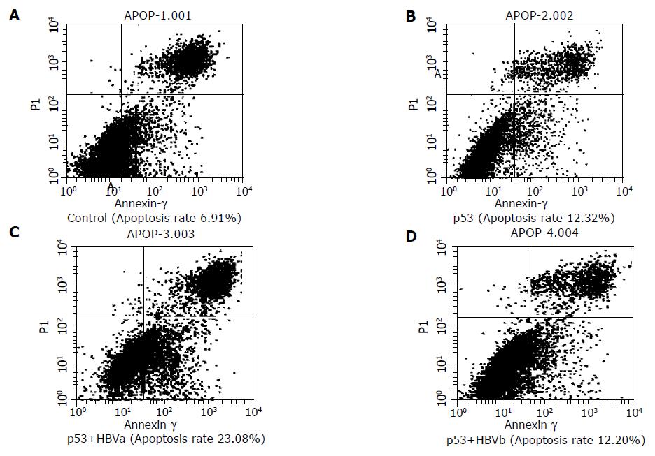
Figure 2 Flow cytometric analysis of cell apoptosis.
SMMU-7721 cells were transfected with p53, p53+HBVa, p53+HBVb. The cells were stained by FITC-labeled annexin V and propidium (A-D).
- Citation: Qu JH, Zhu MH, Lin J, Ni CR, Li FM, Zhu Z, Yu GZ. Effects of hepatitis B virus on p53 expression in hepatoma cell line SMMU-7721. World J Gastroenterol 2005; 11(39): 6212-6215
- URL: https://www.wjgnet.com/1007-9327/full/v11/i39/6212.htm
- DOI: https://dx.doi.org/10.3748/wjg.v11.i39.6212


