Copyright
©The Author(s) 2005.
World J Gastroenterol. Oct 14, 2005; 11(38): 6069-6071
Published online Oct 14, 2005. doi: 10.3748/wjg.v11.i38.6069
Published online Oct 14, 2005. doi: 10.3748/wjg.v11.i38.6069
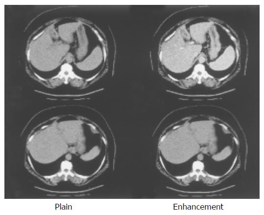
Figure 1 Liver parenchyma is not atrophic.
Thickening of the gall bladder wall is also seen. This finding is compatible with acute hepatitis.
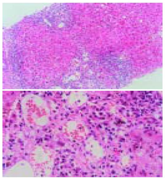
Figure 2 This liver biopsy specimen show the histopathologic appearance of chronic hepatitis.
Interface hepatitis is present with bridging fibrosis, which fibrosis connects portal and cetral arreas. However, dominant plasmacyte infiltrates and rossete formation are not present.
- Citation: Tanaka H, Tujioka H, Ueda H, Hamagami H, Kida Y, Ichinose M. Autoimmune hepatitis triggered by acute hepatitis A. World J Gastroenterol 2005; 11(38): 6069-6071
- URL: https://www.wjgnet.com/1007-9327/full/v11/i38/6069.htm
- DOI: https://dx.doi.org/10.3748/wjg.v11.i38.6069


