Copyright
©The Author(s) 2005.
World J Gastroenterol. Sep 28, 2005; 11(36): 5627-5632
Published online Sep 28, 2005. doi: 10.3748/wjg.v11.i36.5627
Published online Sep 28, 2005. doi: 10.3748/wjg.v11.i36.5627
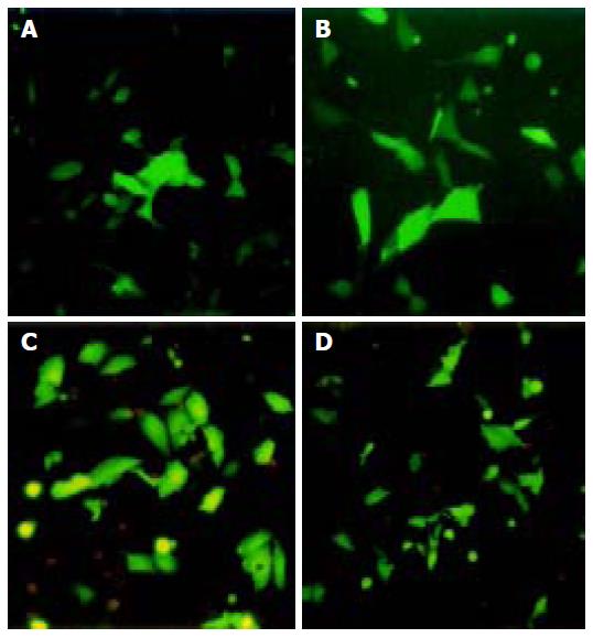
Figure 1 EGFP expressions in HepG2 (A and B) and QBC939 (C and D) cell lines under fluorescence microscope.
After 36 h of transfection, EGFP-expressing cells were visible in group B (A and C) and group C (B and D), but not in group A.
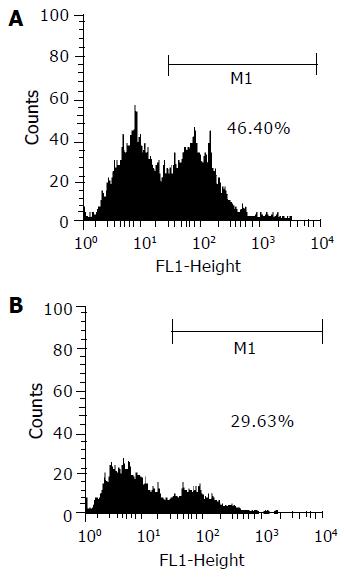
Figure 2 Transfection efficiency evaluated by flow cytometry.
The transfection efficiencies were about 46.4% for HepG2 (A) and 29.6% for QBC939 (B).
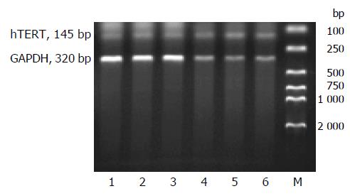
Figure 3 Analysis of hTERT mRNA expression by RT-PCR.
RT-PCR was performed on total RNA extracted from both HepG2 (lanes 1-3) and QBC939 (lanes 4- 6) cell lines transfected with OPTI-MEM medium (lanes 1 and 4), pCMV-X (lanes 2 and 5) and pcDNA3 (lanes 3 and 6) respectively. M: DL 2000 marker.
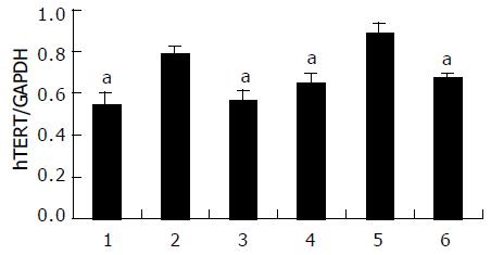
Figure 4 Relative quantitative changes of hTERT mRNA in HepG2 (lanes 1-3) and QBC939 (lanes 4-6) cell lines.
A significant higher expression of hTERT mRNA was observed in both HepG2 and QBC939 cells after being transfected with pCMV-X vector (lanes 2 and 5) compared to those transfected with OPTI-MEM medium (lanes 1 and 4) and pcDNA3 (lanes 3 and 6). aP<0.05 vs others.
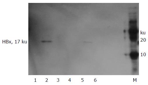
Figure 5 Western blot analysis of HBx protein expression in transfected HepG2 (lanes 1-3) and QBC939 (lanes 4 -6) cell lines.
A marked expression of HBx protein was observed in HepG2 and QBC939 cells when transfected with pCMV -X vector (lanes 2 and 5), but there were no such protein expression in those when transfected with OPTI-MEM medium (lanes 1 and 4) and blank vector (lanes 3 and 6). M: Protein marker.
Figure 6 HBx protein expression in transfected HepG2 cells as assayed by UltraSensitive™ immunocytochemistry (SP×200).
The cells transfected with OPTI-MEM medium (A) and blank vector (C) showed no expression of HBx protein, while some cells transfected with pCMV-X vector (B) showed visible brown positive signals.
Figure 7 HBx protein expression in transfected QBC939 cells as detected by UltraSensitive™ immunocytochemistry (SP×200).
The cells transfected with OPTI-MEM medium (A) and blank vector (C) showed no expression of HBx protein, but some cells transfected with pCMV-X vector (B) showed brown positive signals.
- Citation: Qu ZL, Zou SQ, Cui NQ, Wu XZ, Qin MF, Kong D, Zhou ZL. Upregulation of human telomerase reverse transcriptase mRNA expression by in vitro transfection of hepatitis B virus X gene into human hepatocarcinoma and cholangiocarcinoma cells. World J Gastroenterol 2005; 11(36): 5627-5632
- URL: https://www.wjgnet.com/1007-9327/full/v11/i36/5627.htm
- DOI: https://dx.doi.org/10.3748/wjg.v11.i36.5627


