Copyright
©The Author(s) 2005.
World J Gastroenterol. Sep 28, 2005; 11(36): 5592-5600
Published online Sep 28, 2005. doi: 10.3748/wjg.v11.i36.5592
Published online Sep 28, 2005. doi: 10.3748/wjg.v11.i36.5592
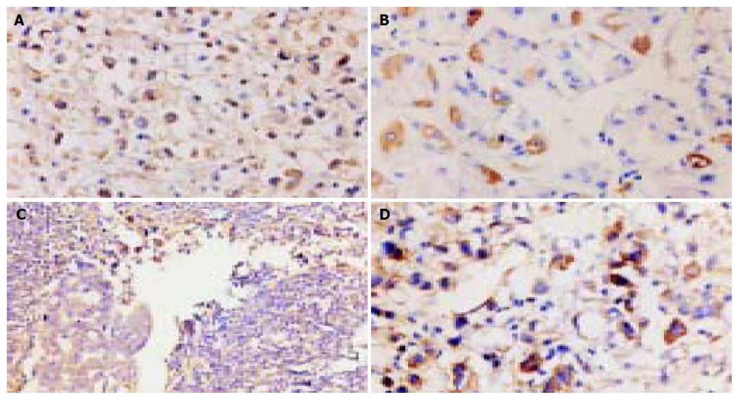
Figure 1 MMP-9 expression in gastric carcinoma.
A: MMP-9 expression showed moderate or diffuse distribution of immunostaining in neoplastic cells (×200); B: single or focal distribution in noncancerous mucosa cells (×400); C: in the invasion front, MMP-9 showed strong positive staining (×200); D: positive staining was also noted in vascular endothelial cells and inflammatory cells (×400).
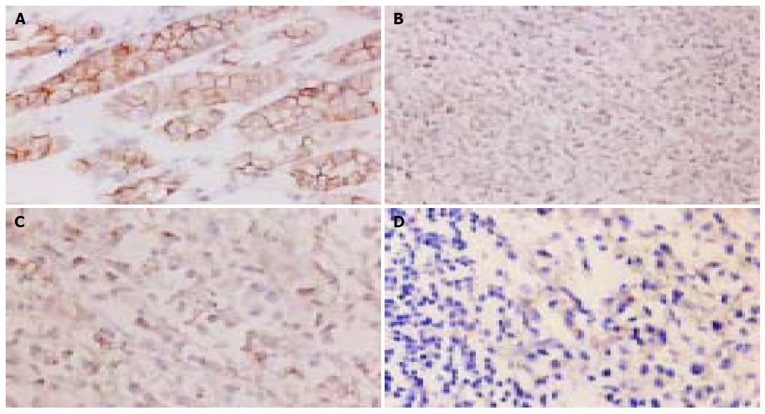
Figure 2 TIMP-2 expression in gastric carcinoma.
A: TIMP-2 expression showed moderate or diffuse distribution in neoplastic cells (×200), B: focal or moderate distribution in noncancerous mucosa cells (×400).
Figure 3 E-cadherin expression in gastric carcinoma.
A: E- cadherin expression showed strong membranous staining in noncancerous mucosa cells, especially at the intercellular border (×400); B-D: abnormal expression was observed in neoplastic tissues which showed reduction or loss of membranous expression and showed cytoplasmic or nuclear staining (B and D, ×200; C, ×400).
Figure 4 Histograms from FCM.
A: A large diploid peak and a smaller tetraploid peak; B: an aneuploid peak (see arrow); C: higher SPF (see arrow).
- Citation: Zhang JF, Zhang YP, Hao FY, Zhang CX, Li YJ, Ji XR. DNA ploidy analysis and expression of MMP-9, TIMP-2, and E-cadherin in gastric carcinoma. World J Gastroenterol 2005; 11(36): 5592-5600
- URL: https://www.wjgnet.com/1007-9327/full/v11/i36/5592.htm
- DOI: https://dx.doi.org/10.3748/wjg.v11.i36.5592


